Copyright
©2011 Baishideng Publishing Group Co.
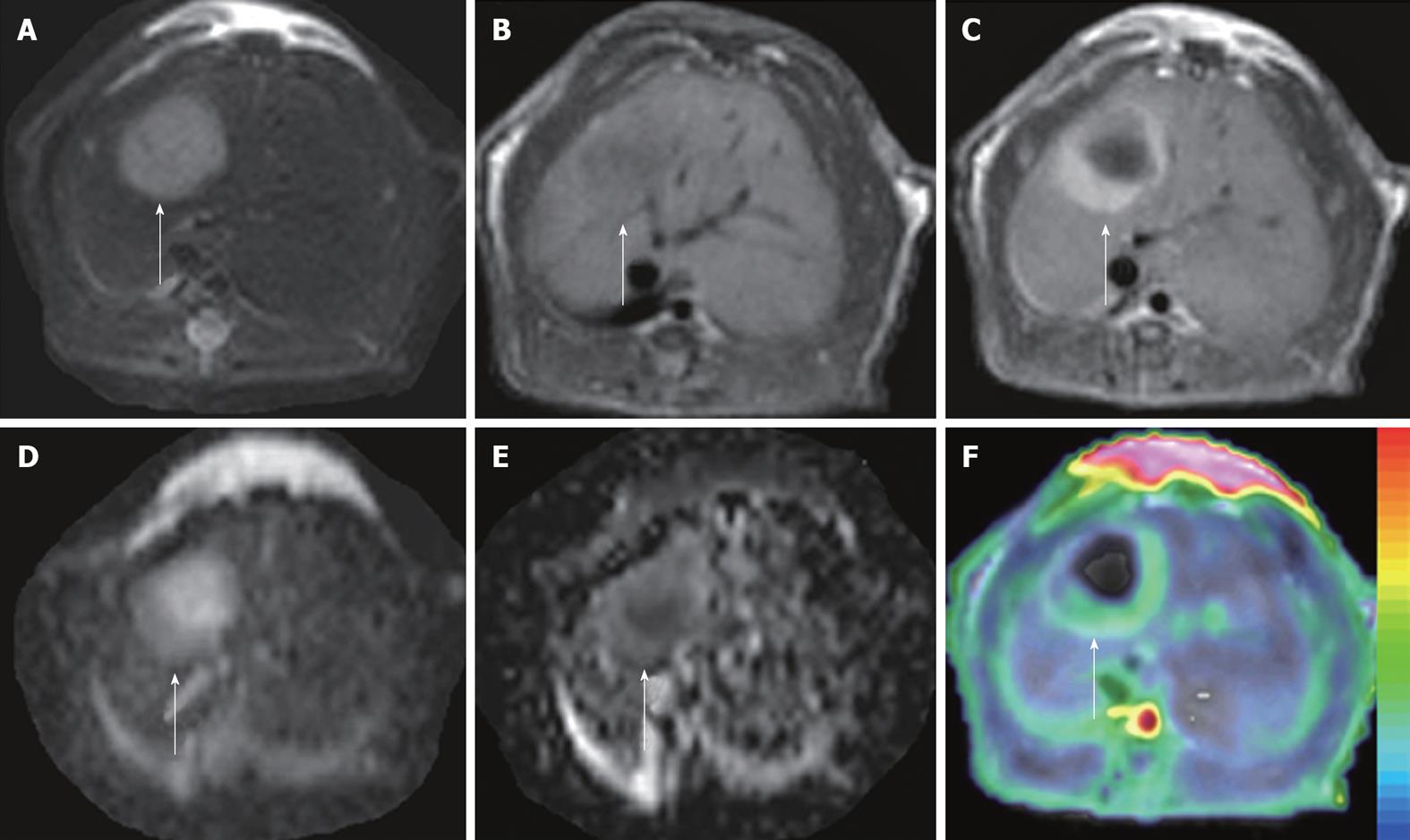
Figure 3 ADCperfusion and initial area under the gadolinium curve in an implanted tumor in rat liver.
At 48 h after iv treatment with CA4P at 10 mg/kg, obvious tumor recurrence with partial recovery of blood supply was demonstrated. The tumor (arrows) appeared hyperintense on T2WI (A) and hypointense on T1WI (B); On CE-T1WI, the tumor relapsed at the periphery, shown as ring enhancement of viable tumor cells (C); ADC10b (derived from 10 b values from 0 to 1000 s/mm2) revealed the hyperintense necrotic center and isointense viable tumor rim (D); On ADCperfusion (ADClow-ADChigh) maps, the relative hyperintensity at the periphery suggested the partial recovery of perfusion, compared to the hypointensity in the necrotic center with perfusion deficit (E); ADCperfusion matched well with CE-T1WI-overlyaed initial area under the gadolinium curve (IAUGC) map (F).
- Citation: Wang H, Marchal G, Ni Y. Multiparametric MRI biomarkers for measuring vascular disrupting effect on cancer. World J Radiol 2011; 3(1): 1-16
- URL: https://www.wjgnet.com/1949-8470/full/v3/i1/1.htm
- DOI: https://dx.doi.org/10.4329/wjr.v3.i1.1


