Copyright
©2010 Baishideng Publishing Group Co.
World J Radiol. Sep 28, 2010; 2(9): 345-353
Published online Sep 28, 2010. doi: 10.4329/wjr.v2.i9.345
Published online Sep 28, 2010. doi: 10.4329/wjr.v2.i9.345
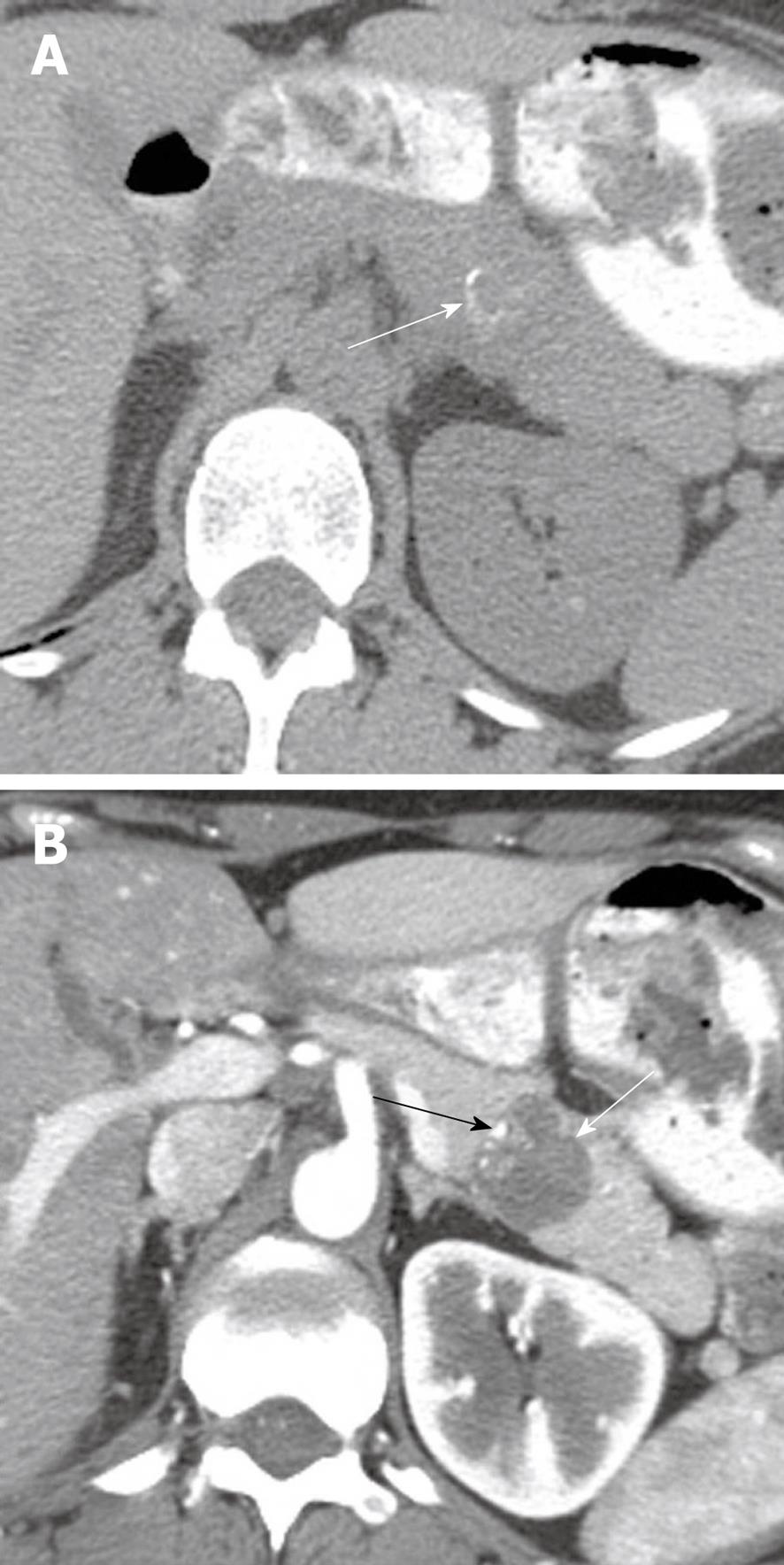
Figure 8 A 30-year-old woman with increasing abdominal discomfort and bloating.
A: Axial non contrast computed tomography (CT) of the abdomen shows a solid mass in the pancreatic tail containing curvilinear calcification (arrow); B: Axial contrast-enhanced CT of the abdomen shows a hypoattenuating mass (white arrow) in the pancreas containing eccentric calcifications (black arrow) consistent with a solid papillary epithelial neoplasm.
- Citation: Bhosale P, Balachandran A, Tamm E. Imaging of benign and malignant cystic pancreatic lesions and a strategy for follow up. World J Radiol 2010; 2(9): 345-353
- URL: https://www.wjgnet.com/1949-8470/full/v2/i9/345.htm
- DOI: https://dx.doi.org/10.4329/wjr.v2.i9.345


