Copyright
©2010 Baishideng Publishing Group Co.
World J Radiol. Sep 28, 2010; 2(9): 345-353
Published online Sep 28, 2010. doi: 10.4329/wjr.v2.i9.345
Published online Sep 28, 2010. doi: 10.4329/wjr.v2.i9.345
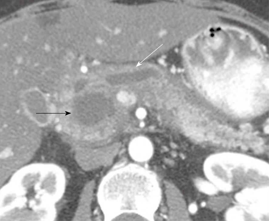
Figure 1 A 49-year-old woman with a history of epigastric and lower abdominal pain accompanied by abnormal liver function studies.
Axial contrast-enhanced computed tomography scan of the abdomen shows cystic change (black arrow) within the pancreas with associated biliary ductal dilation (white arrow). When correlated with the patient’s history and endoscopic ultrasound with FNA findings, this was consistent with a pseudocyst.
- Citation: Bhosale P, Balachandran A, Tamm E. Imaging of benign and malignant cystic pancreatic lesions and a strategy for follow up. World J Radiol 2010; 2(9): 345-353
- URL: https://www.wjgnet.com/1949-8470/full/v2/i9/345.htm
- DOI: https://dx.doi.org/10.4329/wjr.v2.i9.345


