
















©2010 Baishideng Publishing Group Co.
World J Radiol. Aug 28, 2010; 2(8): 334-338
Published online Aug 28, 2010. doi: 10.4329/wjr.v2.i8.334
Published online Aug 28, 2010. doi: 10.4329/wjr.v2.i8.334
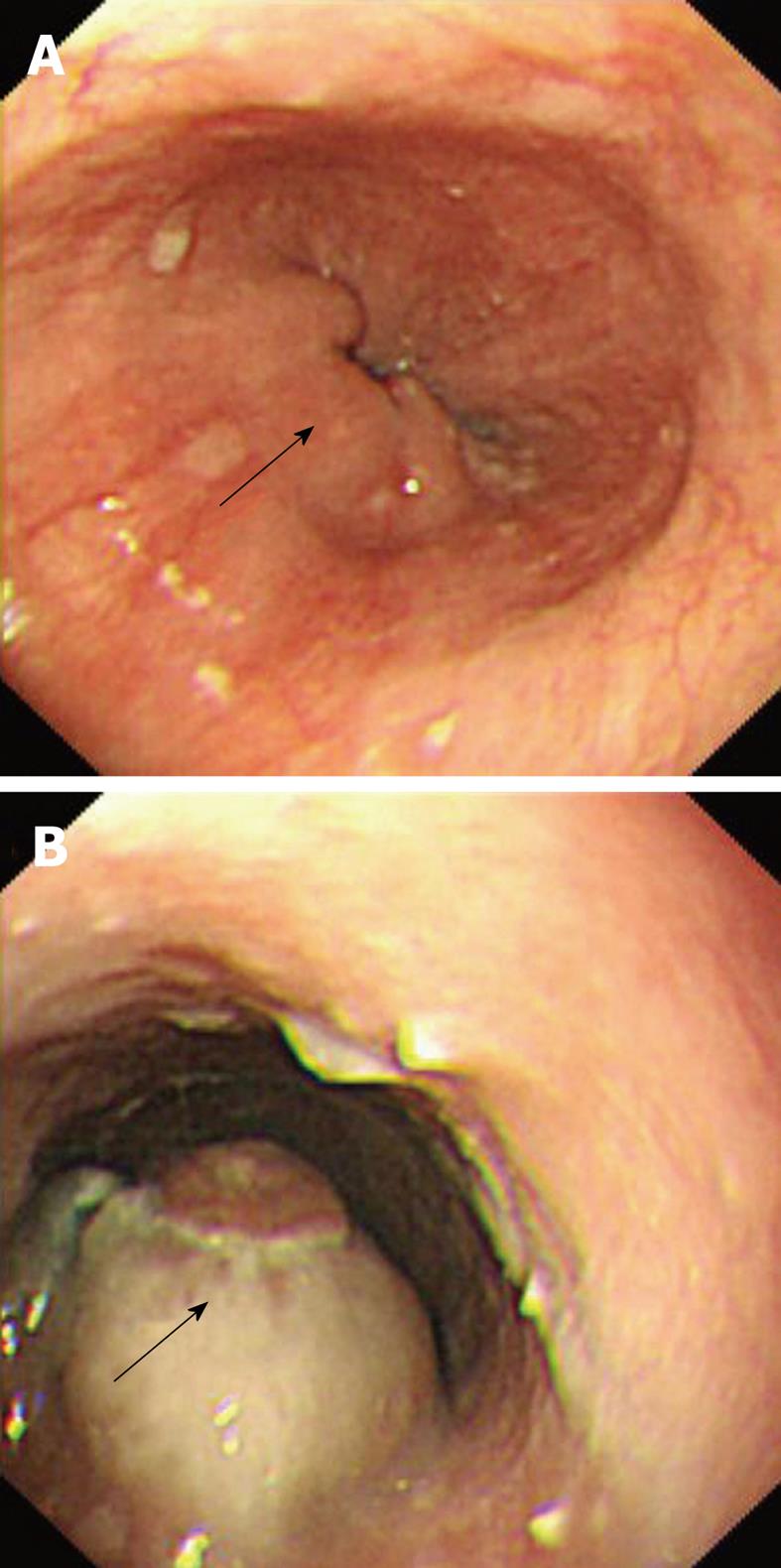
Figure 4 Esophagogastroduodenoscopy (A and B) showing multiple solid, firm nodular lesions in the lower esophagus.
Overlying mucosa appearing relatively normal (black arrows).
- Citation: Ghimire P, Wu GY, Zhu L. Primary esophageal lymphoma in immunocompetent patients: Two case reports and literature review. World J Radiol 2010; 2(8): 334-338
- URL: https://www.wjgnet.com/1949-8470/full/v2/i8/334.htm
- DOI: https://dx.doi.org/10.4329/wjr.v2.i8.334