
















©2010 Baishideng Publishing Group Co.
World J Radiol. Aug 28, 2010; 2(8): 298-308
Published online Aug 28, 2010. doi: 10.4329/wjr.v2.i8.298
Published online Aug 28, 2010. doi: 10.4329/wjr.v2.i8.298
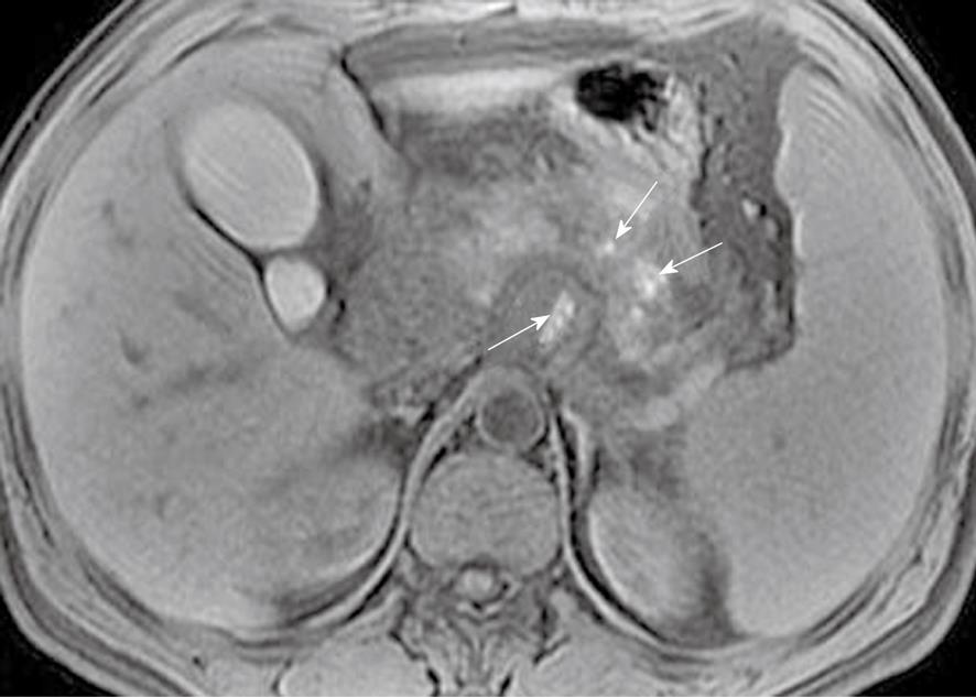
Figure 7 Acute pancreatitis in a 30-year-old man.
Axial non-enhanced magnetic resonance T1-weighted with fat-suppression image obtained at the time of hospital admission shows multiple patchy hemorrhagic foci (like “salt”) (arrows) in the pancreas and in the fatty tissue behind the pancreas.
- Citation: Xiao B, Zhang XM. Magnetic resonance imaging for acute pancreatitis. World J Radiol 2010; 2(8): 298-308
- URL: https://www.wjgnet.com/1949-8470/full/v2/i8/298.htm
- DOI: https://dx.doi.org/10.4329/wjr.v2.i8.298