Copyright
©2010 Baishideng Publishing Group Co.
World J Radiol. Aug 28, 2010; 2(8): 289-297
Published online Aug 28, 2010. doi: 10.4329/wjr.v2.i8.289
Published online Aug 28, 2010. doi: 10.4329/wjr.v2.i8.289
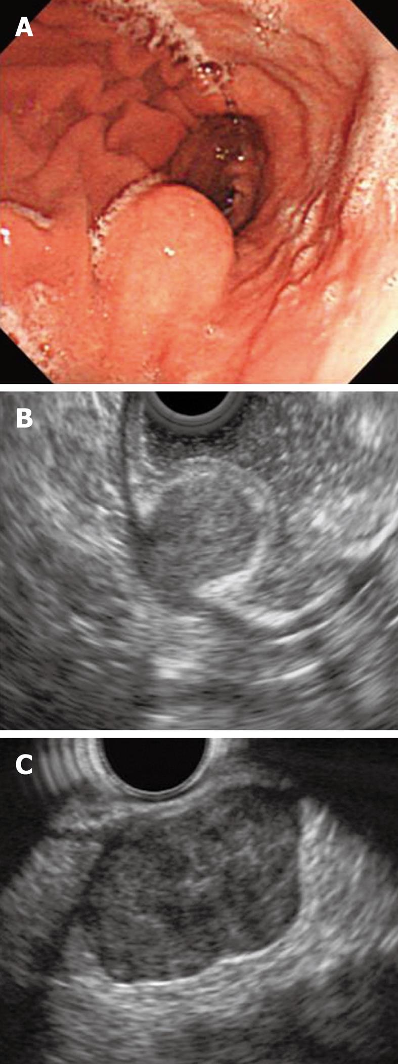
Figure 6 Endoscopic and endoscopic ultrasonography finding of gastrointestinal stromal tumor.
A: Endoscopic view of subepithelial lesion of the posterior side of the greater curvature of the gastric body; B: Endoscopic ultrasonography shows hypoechoic, homogeneous 2 cm tumor developed within the 4th layer (low risk gastrointestinal stromal tumor of the stomach); C: 35 mm hypoechoic, heterogeneous, lobulated submucosal lesion with exogastric growth developed within the 4th layer (high risk gastrointestinal stromal tumor of the stomach).
- Citation: Sakamoto H, Kitano M, Kudo M. Diagnosis of subepithelial tumors in the upper gastrointestinal tract by endoscopic ultrasonography. World J Radiol 2010; 2(8): 289-297
- URL: https://www.wjgnet.com/1949-8470/full/v2/i8/289.htm
- DOI: https://dx.doi.org/10.4329/wjr.v2.i8.289


