
















©2010 Baishideng Publishing Group Co.
World J Radiol. Jul 28, 2010; 2(7): 237-248
Published online Jul 28, 2010. doi: 10.4329/wjr.v2.i7.237
Published online Jul 28, 2010. doi: 10.4329/wjr.v2.i7.237
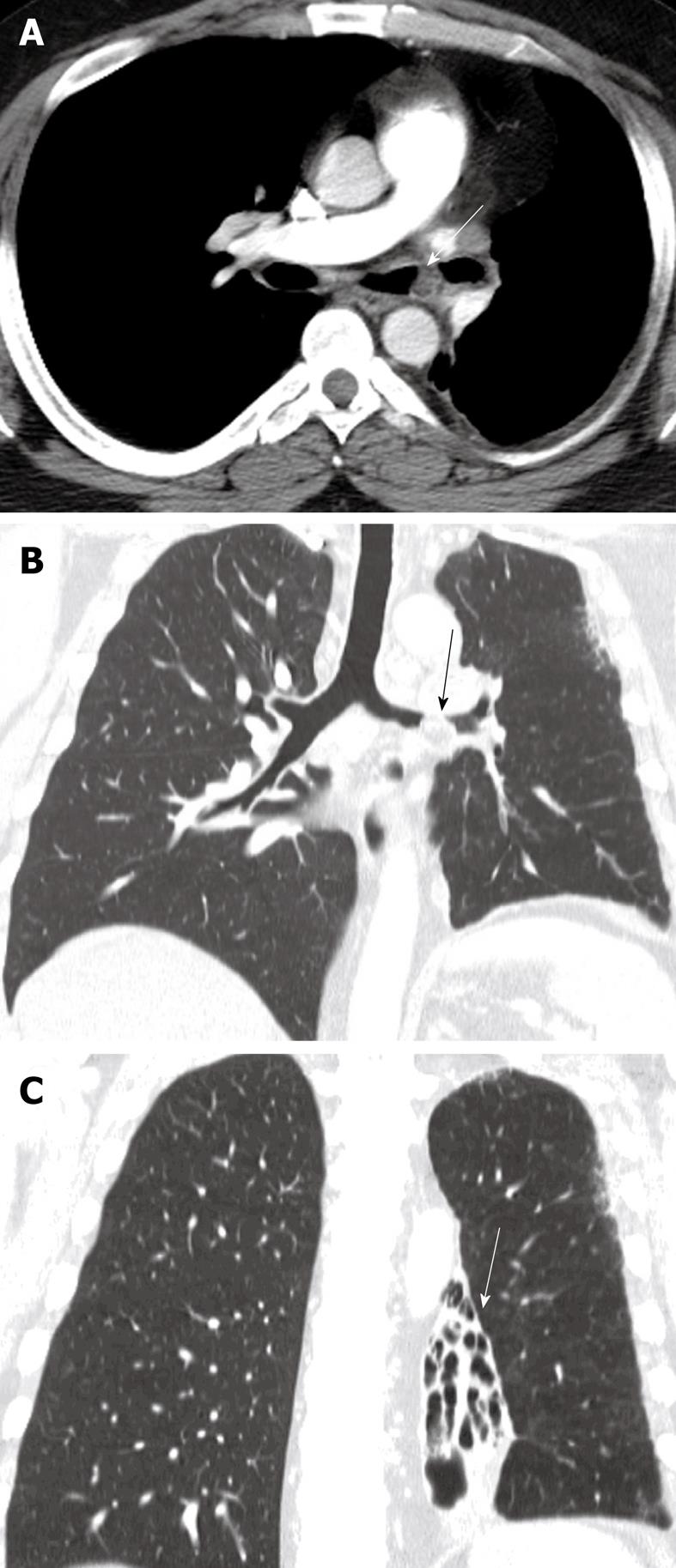
Figure 16 Endobronchial hamartoma.
Axial computed tomography image (A) and the multiplanar reconstruction in the lung window (B and C) shows chronic bronchiectasis in the left lower lobe (arrow) secondary to a long-standing slow-growing obstruction of the left main bronchus and recurrent infections (arrows).
- Citation: Laroia AT, Thompson BH, Laroia ST, Beek EJV. Modern imaging of the tracheo-bronchial tree. World J Radiol 2010; 2(7): 237-248
- URL: https://www.wjgnet.com/1949-8470/full/v2/i7/237.htm
- DOI: https://dx.doi.org/10.4329/wjr.v2.i7.237