Copyright
©2010 Baishideng Publishing Group Co.
World J Radiol. May 28, 2010; 2(5): 159-165
Published online May 28, 2010. doi: 10.4329/wjr.v2.i5.159
Published online May 28, 2010. doi: 10.4329/wjr.v2.i5.159
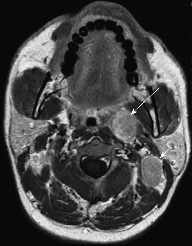
Figure 11 Axial post-contrast T1 weighted MRI in a patient with nasopharyngeal carcinoma showing an enlarged left sided metastatic retropharyngeal node deep to the oropharynx (long arrow).
An enlarged metastatic left upper internal jugular node posterior to the left internal jugular vein (level IIB) is also present which is a very common site of nodal metastases (short arrow).
- Citation: King AD, Bhatia KSS. Magnetic resonance imaging staging of nasopharyngeal carcinoma in the head and neck. World J Radiol 2010; 2(5): 159-165
- URL: https://www.wjgnet.com/1949-8470/full/v2/i5/159.htm
- DOI: https://dx.doi.org/10.4329/wjr.v2.i5.159


