Copyright
©2010 Baishideng Publishing Group Co.
World J Radiol. May 28, 2010; 2(5): 159-165
Published online May 28, 2010. doi: 10.4329/wjr.v2.i5.159
Published online May 28, 2010. doi: 10.4329/wjr.v2.i5.159
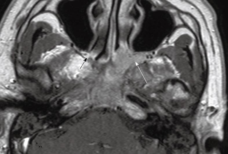
Figure 8 Axial post-contrast T1 weighted MRI showing contiguous extension of nasopharyngeal carcinoma into the left pterygopalatine fossa which is expanded (long arrow).
Compare this with the normal hyperintense fat signal in the narrow pterygopalatine fossa on the contralateral side (short arrow).
- Citation: King AD, Bhatia KSS. Magnetic resonance imaging staging of nasopharyngeal carcinoma in the head and neck. World J Radiol 2010; 2(5): 159-165
- URL: https://www.wjgnet.com/1949-8470/full/v2/i5/159.htm
- DOI: https://dx.doi.org/10.4329/wjr.v2.i5.159


