Copyright
©2010 Baishideng Publishing Group Co.
World J Radiol. Apr 28, 2010; 2(4): 122-134
Published online Apr 28, 2010. doi: 10.4329/wjr.v2.i4.122
Published online Apr 28, 2010. doi: 10.4329/wjr.v2.i4.122
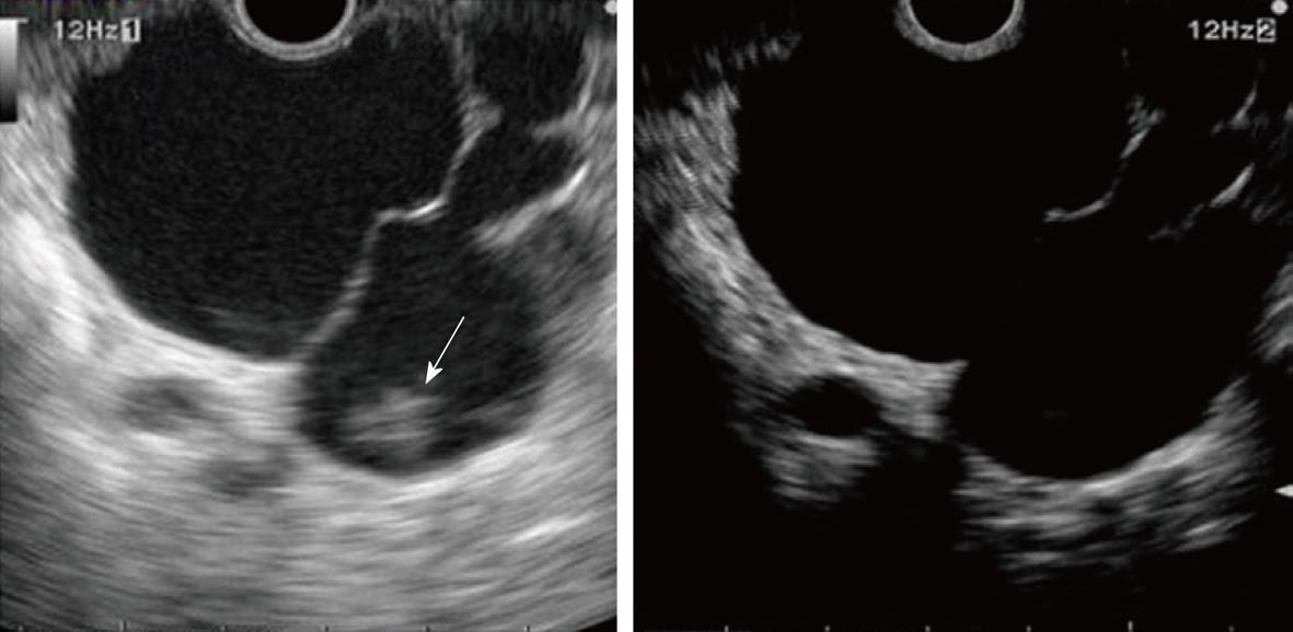
Figure 12 IPMN of side branch type.
Left (B-mode image): The nodule (arrow) in dilatation of the side branch cannot be distinguished between sediment and tumor by B-EUS; Right (contrast image): Contrast-enhanced harmonic EUS reveals that this nodule is sediment.
- Citation: Sakamoto H, Kitano M, Kamata K, El-Masry M, Kudo M. Diagnosis of pancreatic tumors by endoscopic ultrasonography. World J Radiol 2010; 2(4): 122-134
- URL: https://www.wjgnet.com/1949-8470/full/v2/i4/122.htm
- DOI: https://dx.doi.org/10.4329/wjr.v2.i4.122


