Copyright
©2010 Baishideng Publishing Group Co.
World J Radiol. Apr 28, 2010; 2(4): 122-134
Published online Apr 28, 2010. doi: 10.4329/wjr.v2.i4.122
Published online Apr 28, 2010. doi: 10.4329/wjr.v2.i4.122
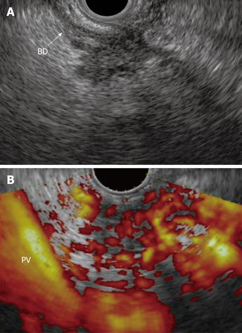
Figure 10 Focal chronic pancreatitis.
A: EUS shows a mass with an irregular and inhomogeneous echo pattern at the head of the pancreas; B: Contrast-enhanced power Doppler EUS shows an isovascular nodule compared with the surrounding pancreatic tissue. BD: Bile duct; PV: Portal vein.
- Citation: Sakamoto H, Kitano M, Kamata K, El-Masry M, Kudo M. Diagnosis of pancreatic tumors by endoscopic ultrasonography. World J Radiol 2010; 2(4): 122-134
- URL: https://www.wjgnet.com/1949-8470/full/v2/i4/122.htm
- DOI: https://dx.doi.org/10.4329/wjr.v2.i4.122


