Copyright
©2010 Baishideng Publishing Group Co.
World J Radiol. Apr 28, 2010; 2(4): 122-134
Published online Apr 28, 2010. doi: 10.4329/wjr.v2.i4.122
Published online Apr 28, 2010. doi: 10.4329/wjr.v2.i4.122
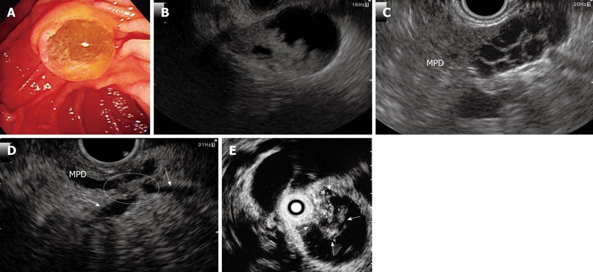
Figure 8 Intraductal papillary mucinous neoplasms (IPMN).
A: An endoscopic diagnosis of an IPMN can be established if the “fish-eye” ampulla is visualized in minority cases; B: IPMN of main duct type. EUS shows a mural nodule within by the mucinous dilatation of the pancreatic ducts, with involvement of the main duct at the tail of the pancreas; C: IPMN of side branch type. EUS shows a multiple dilatation of the side branch at the neck of the pancreas; D: IPMN of the combined type. EUS show a mural nodule stretching (circle) over the main pancreatic duct and side branches (arrows) at the body of the pancreas. E: IPMN of main duct type. Intraductal ultrasonography (IDUS) can identify tumor nodule development into the main pancreatic duct (arrows). MPD: Main pancreatic duct.
- Citation: Sakamoto H, Kitano M, Kamata K, El-Masry M, Kudo M. Diagnosis of pancreatic tumors by endoscopic ultrasonography. World J Radiol 2010; 2(4): 122-134
- URL: https://www.wjgnet.com/1949-8470/full/v2/i4/122.htm
- DOI: https://dx.doi.org/10.4329/wjr.v2.i4.122


