
















©2010 Baishideng.
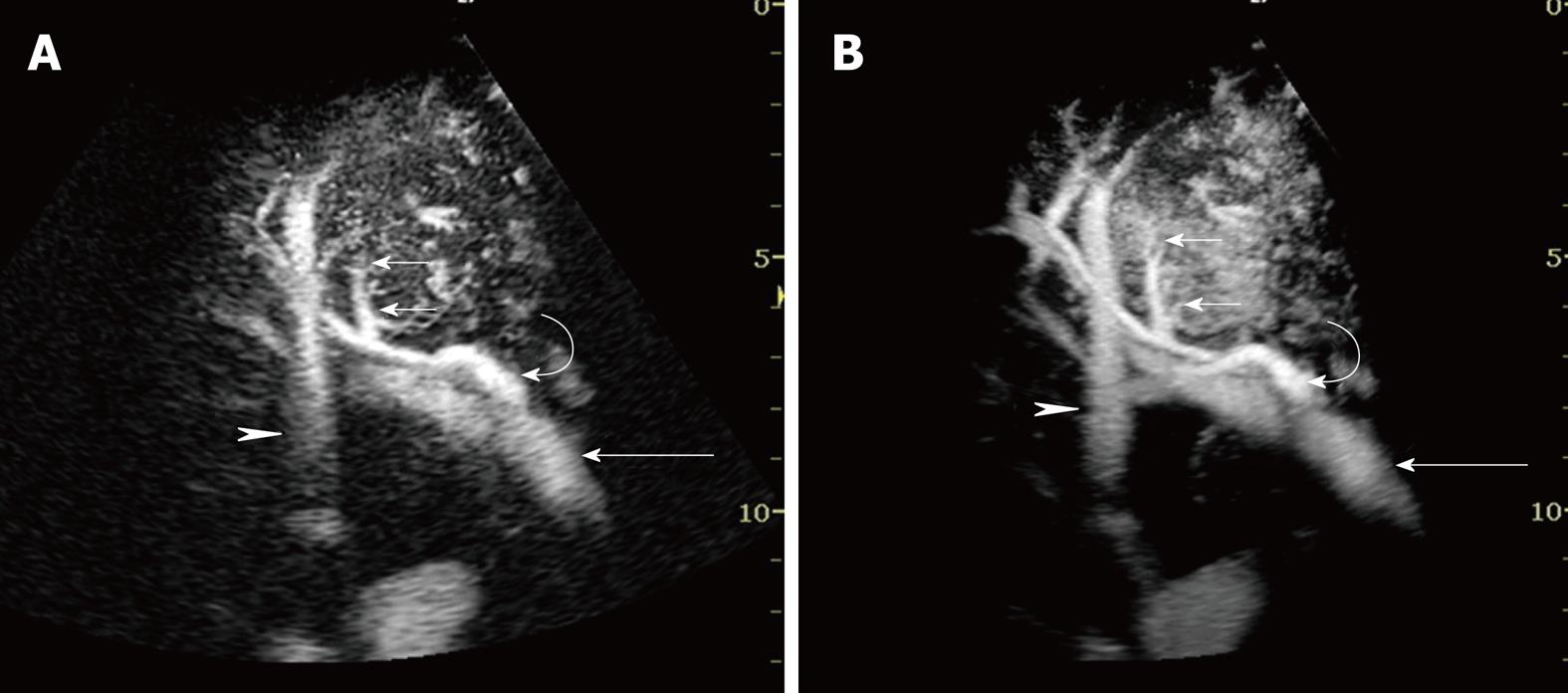
Figure 2 A 76-year-old man with HCC (maximum diameter 40 mm) in segment VIII.
A: Early phase Sonazoid-enhanced US by CHA mode at a high MI shows intratumoral vessels (small arrows), right portal vein (arrow), hepatic artery (curved arrow), and hepatic vein (arrowhead); B: Accumulation maximum intensity holding image in the early phase. Sonazoid-enhanced US by CHA mode at a high MI more clearly shows the serial images of intratumoral vessels (small arrows), right portal vein (arrow), hepatic artery (curved arrow), and hepatic vein (arrowhead) than the images in A.
- Citation: Numata K, Luo W, Morimoto M, Kondo M, Kunishi Y, Sasaki T, Nozaki A, Tanaka K. Contrast enhanced ultrasound of hepatocellular carcinoma. World J Radiol 2010; 2(2): 68-82
- URL: https://www.wjgnet.com/1949-8470/full/v2/i2/68.htm
- DOI: https://dx.doi.org/10.4329/wjr.v2.i2.68