Copyright
©2010 Baishideng Publishing Group Co.
World J Radiol. Nov 28, 2010; 2(11): 440-448
Published online Nov 28, 2010. doi: 10.4329/wjr.v2.i11.440
Published online Nov 28, 2010. doi: 10.4329/wjr.v2.i11.440
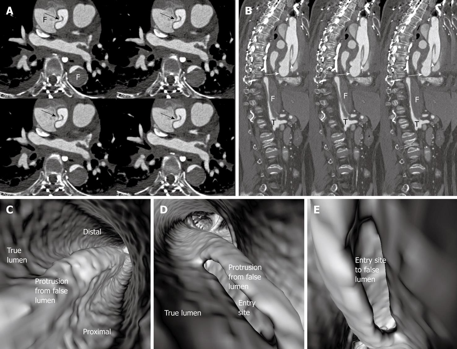
Figure 6 Stanford type A dissection with direct communication between the true lumen and false lumens (arrows in A).
The true lumen (T) was obviously narrowed due to compression by the false lumen (F) with thrombus formed in the false lumen as shown on the sagittal reformatted image (B). The intimal flap (short arrows in B) arises from the level of the left ventricle (long arrows in B); C: A protrusion sign was observed on virtual intravascular endoscopy (VIE) images; D, E: The long intimal tear was identified at the ascending aorta posterior to the protrusion from the false lumen on different VIE visualizations. Pleural effusion is present at both sides on 2D axial images.
- Citation: Sun Z, Cao Y. Multislice CT virtual intravascular endoscopy of aortic dissection: A pictorial essay. World J Radiol 2010; 2(11): 440-448
- URL: https://www.wjgnet.com/1949-8470/full/v2/i11/440.htm
- DOI: https://dx.doi.org/10.4329/wjr.v2.i11.440


