Copyright
©2010 Baishideng Publishing Group Co.
World J Radiol. Nov 28, 2010; 2(11): 440-448
Published online Nov 28, 2010. doi: 10.4329/wjr.v2.i11.440
Published online Nov 28, 2010. doi: 10.4329/wjr.v2.i11.440
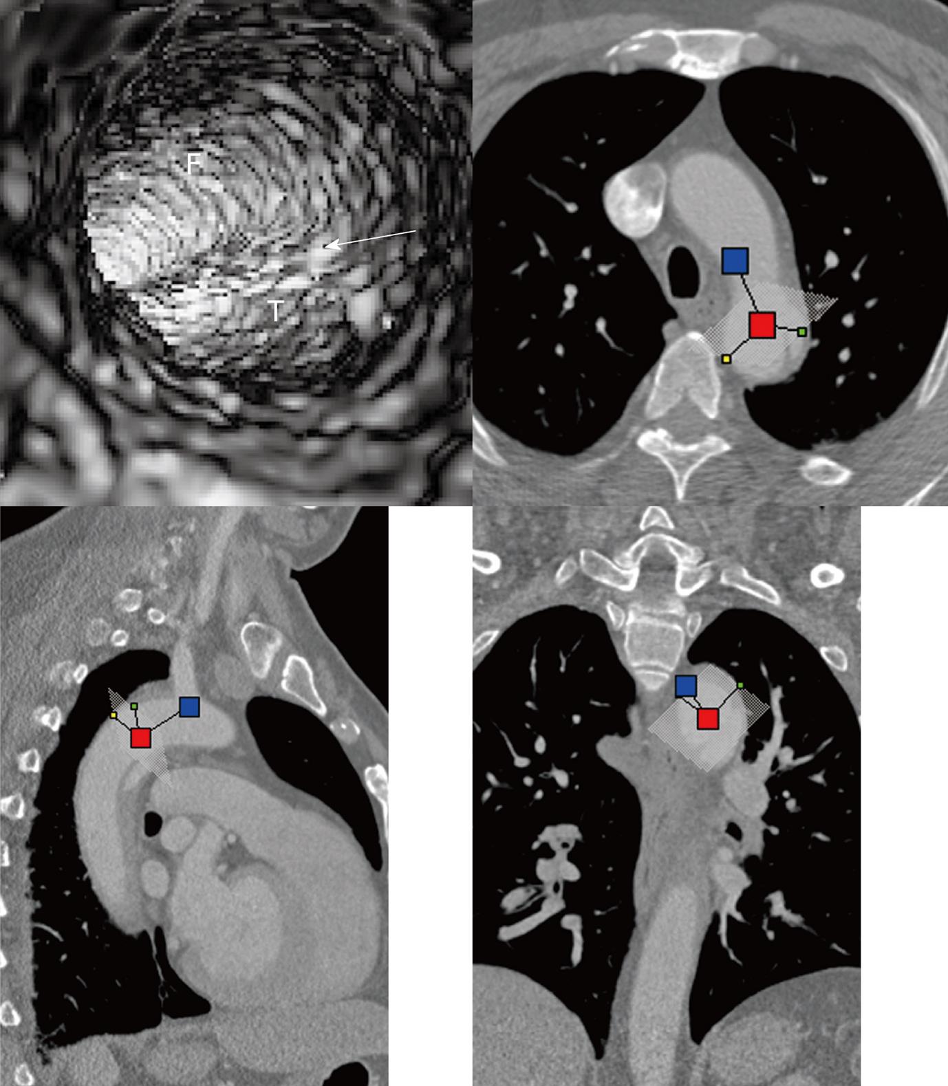
Figure 1 Virtual intravascular endoscopy visualization of the true lumen, false lumen and intimal flap (arrow) in a Stanford type B aortic dissection.
Corresponding orthogonal views (axial, coronal and sagittal) confirm the location of virtual intravascular endoscopy position inside the false lumen (F) looking toward the true lumen (T) and intimal flap.
- Citation: Sun Z, Cao Y. Multislice CT virtual intravascular endoscopy of aortic dissection: A pictorial essay. World J Radiol 2010; 2(11): 440-448
- URL: https://www.wjgnet.com/1949-8470/full/v2/i11/440.htm
- DOI: https://dx.doi.org/10.4329/wjr.v2.i11.440


