
















©2010 Baishideng Publishing Group Co.
World J Radiol. Oct 28, 2010; 2(10): 405-409
Published online Oct 28, 2010. doi: 10.4329/wjr.v2.i10.405
Published online Oct 28, 2010. doi: 10.4329/wjr.v2.i10.405
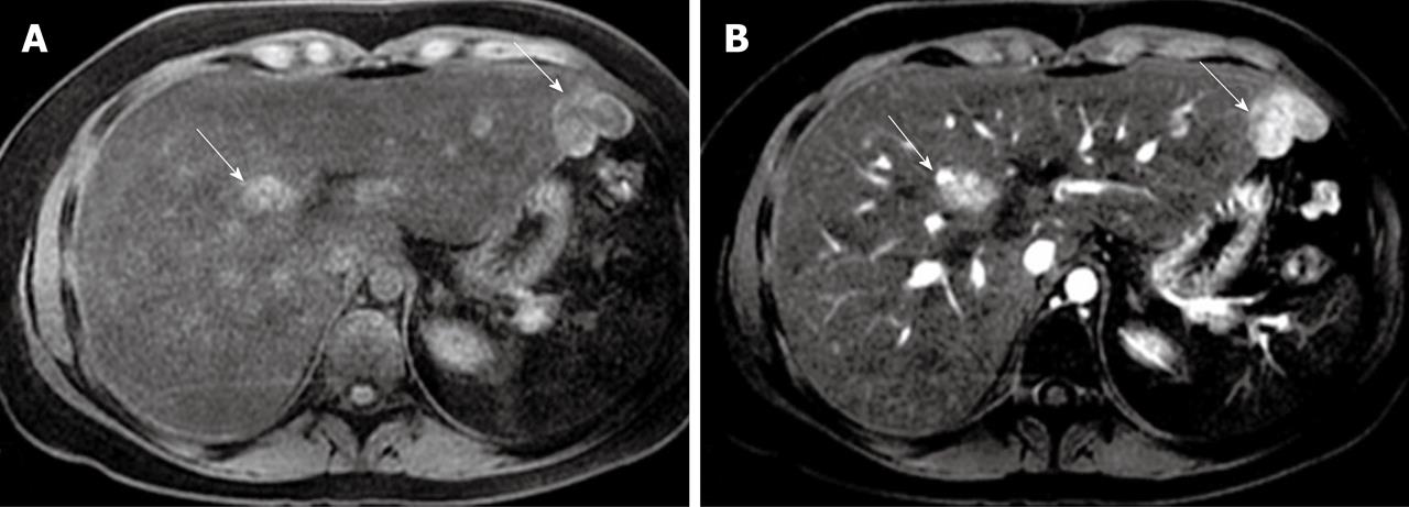
Figure 2 Axial pre-contrast T1-weighted magnetic resonance imaging demonstrating focal nodular hyperplasia lesions with an increased intensity (arrows) (A) and enhanced focal nodular hyperplasia lesions at the arterial phase (arrows) (B).
- Citation: Kayhan A, Venu N, Lakadamyalı H, Jensen D, Oto A. Multiple progressive focal nodular hyperplasia lesions of liver in a patient with hemosiderosis. World J Radiol 2010; 2(10): 405-409
- URL: https://www.wjgnet.com/1949-8470/full/v2/i10/405.htm
- DOI: https://dx.doi.org/10.4329/wjr.v2.i10.405