Copyright
©2010 Baishideng Publishing Group Co.
World J Radiol. Oct 28, 2010; 2(10): 384-398
Published online Oct 28, 2010. doi: 10.4329/wjr.v2.i10.384
Published online Oct 28, 2010. doi: 10.4329/wjr.v2.i10.384
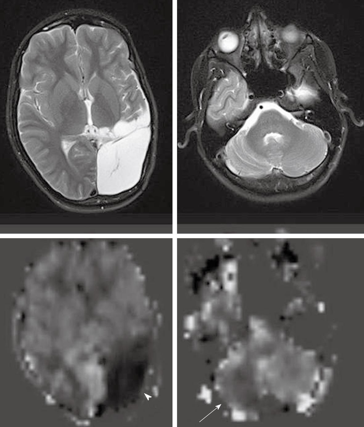
Figure 14 A case of old infarction with encephalomalacic change in left cerebral hemisphere.
Arterial spin labeling cerebral blood flow maps (bottom row) demonstrate no perfusion to the encephalomalacic brain (white arrowhead) and decreased perfusion in the mildly atrophic right cerebellar hemisphere (white arrow), consistent with cross cerebellar diaschisis.
- Citation: Petcharunpaisan S, Ramalho J, Castillo M. Arterial spin labeling in neuroimaging. World J Radiol 2010; 2(10): 384-398
- URL: https://www.wjgnet.com/1949-8470/full/v2/i10/384.htm
- DOI: https://dx.doi.org/10.4329/wjr.v2.i10.384


