Copyright
©2010 Baishideng Publishing Group Co.
World J Radiol. Oct 28, 2010; 2(10): 384-398
Published online Oct 28, 2010. doi: 10.4329/wjr.v2.i10.384
Published online Oct 28, 2010. doi: 10.4329/wjr.v2.i10.384
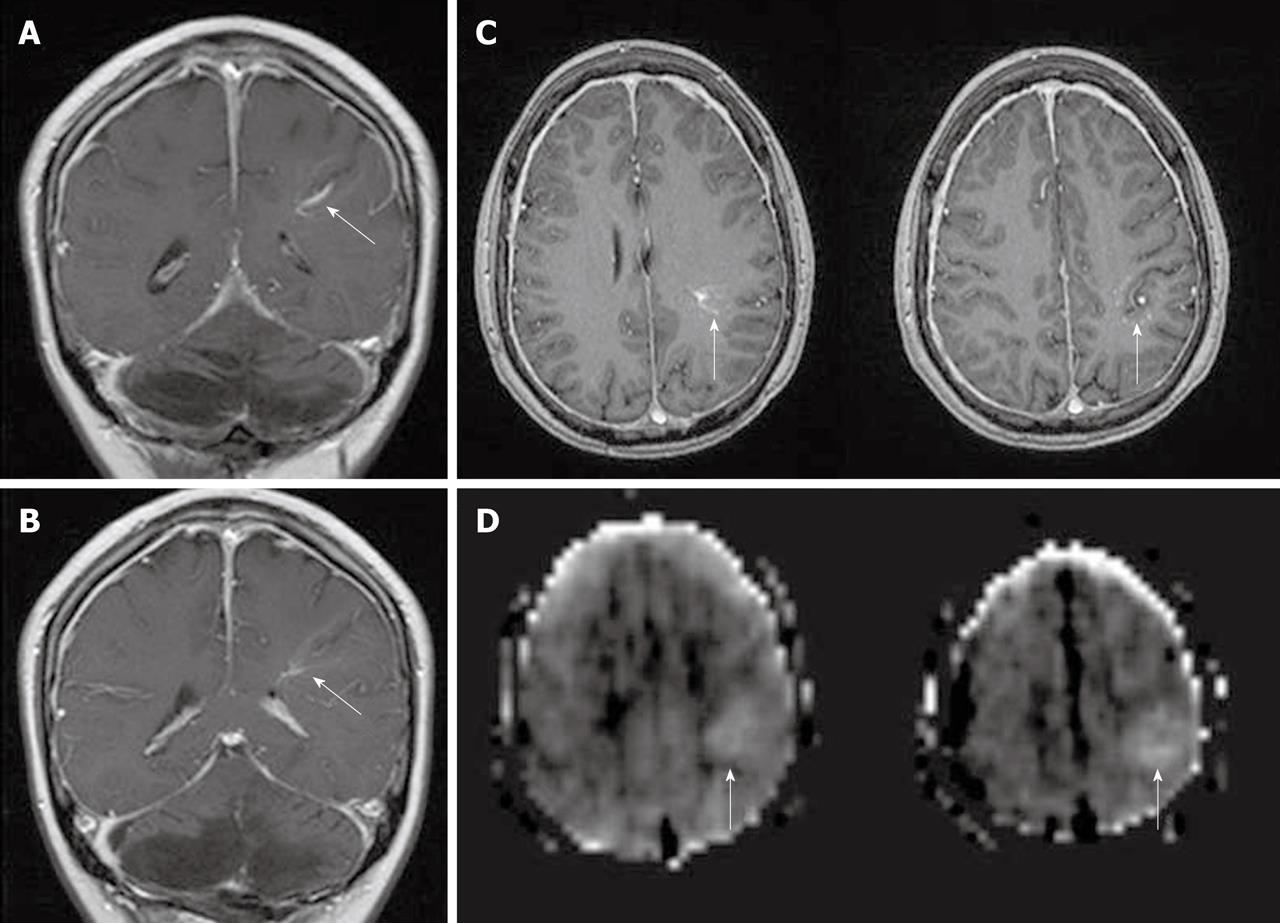
Figure 11 Developmental venous anomaly in the left frontoparietal lobe, seen as enhancing dilated medullary veins draining into the transcortical collector vein on coronal (A and B) and axial (C) post gadolinium T1WI (white arrows).
Arterial spin labeling cerebral blood flow maps (D) demonstrate hyperperfusion in the developmental venous anomaly and surrounding brain parenchyma (white arrows).
- Citation: Petcharunpaisan S, Ramalho J, Castillo M. Arterial spin labeling in neuroimaging. World J Radiol 2010; 2(10): 384-398
- URL: https://www.wjgnet.com/1949-8470/full/v2/i10/384.htm
- DOI: https://dx.doi.org/10.4329/wjr.v2.i10.384


