Copyright
©2010 Baishideng Publishing Group Co.
World J Radiol. Oct 28, 2010; 2(10): 384-398
Published online Oct 28, 2010. doi: 10.4329/wjr.v2.i10.384
Published online Oct 28, 2010. doi: 10.4329/wjr.v2.i10.384
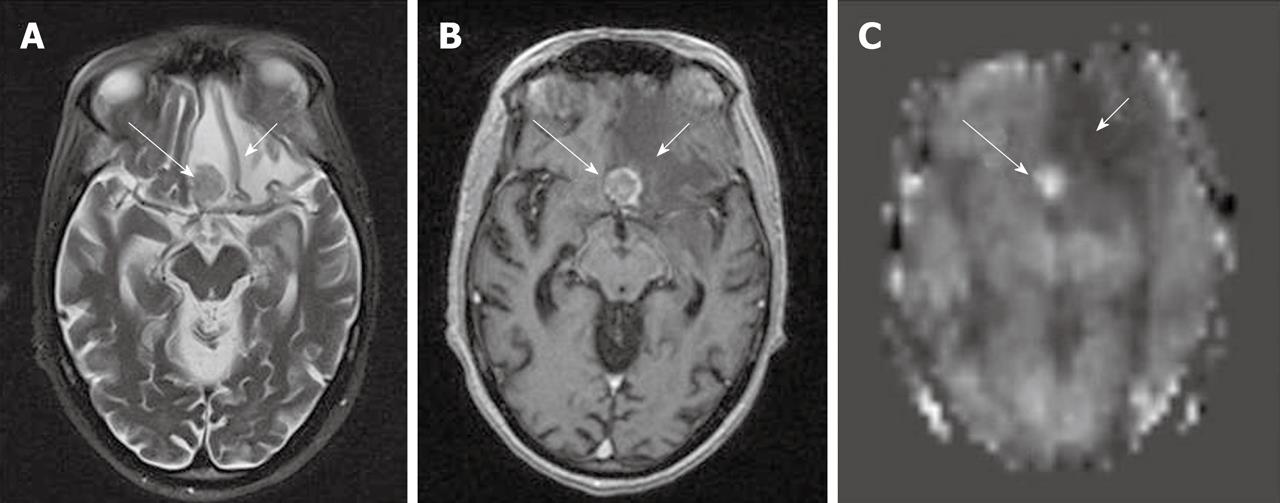
Figure 8 A case of non-small cell lung cancer with left frontal metastases.
The lesion shows low to isosignal intensity on T2WI (A), peripheral and central enhancement on post gadolinium T1WI (B) and hyperperfusion on arterial spin labeling (ASL) cerebral blood flow (CBF) map (C) (long white arrows). The area of perilesional edema (short white arrows) also shows hypoperfusion on ASL CBF map.
- Citation: Petcharunpaisan S, Ramalho J, Castillo M. Arterial spin labeling in neuroimaging. World J Radiol 2010; 2(10): 384-398
- URL: https://www.wjgnet.com/1949-8470/full/v2/i10/384.htm
- DOI: https://dx.doi.org/10.4329/wjr.v2.i10.384


