
















Copyright: ©Author(s) 2026.
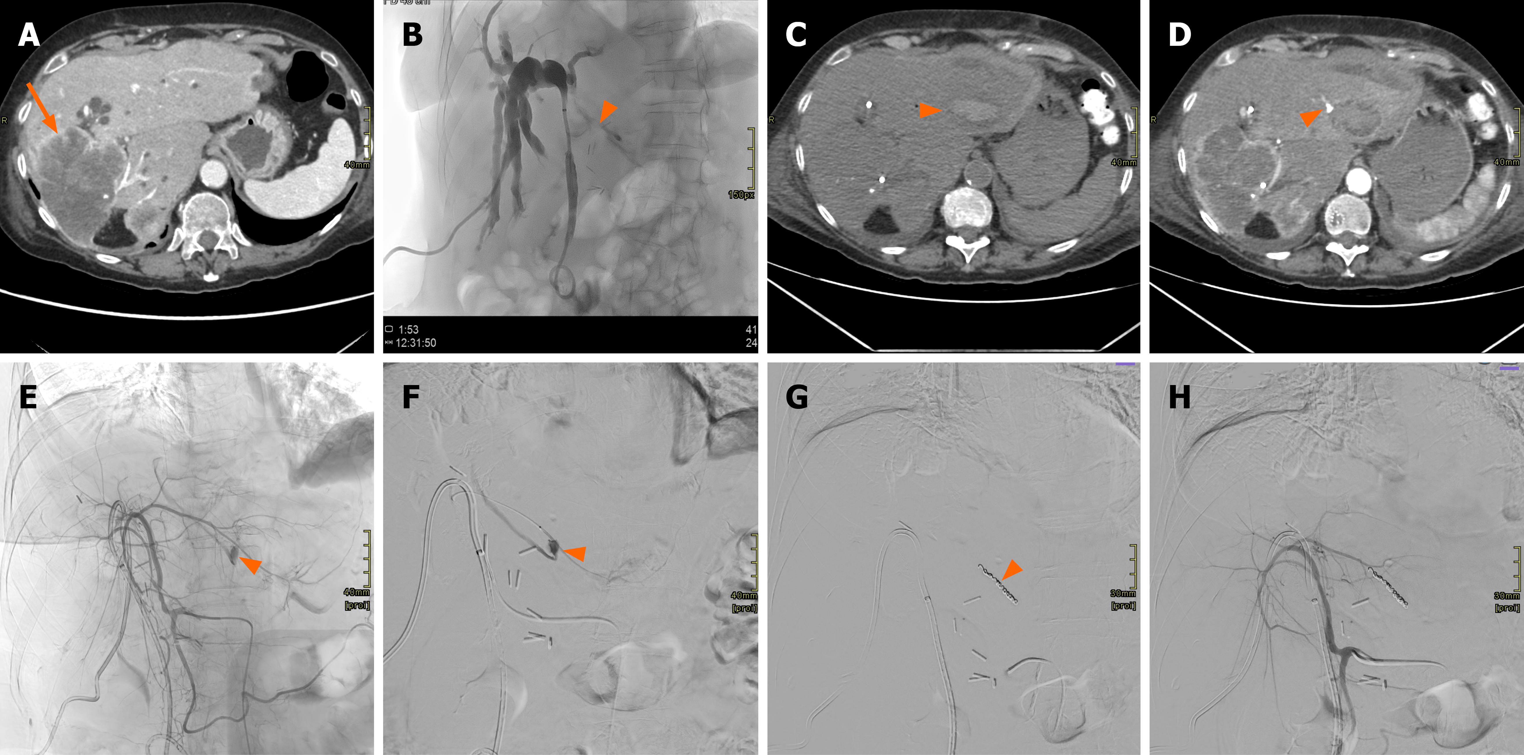
Figure 7 Patient with recurrent cholangiocarcinoma involving segment IV, approximately 18 months after prior right hepatectomy.
A: Computed tomography scan showing a large mass (arrow) in the right liver lobe, in keeping with cholangiocarcinoma; B: The patient underwent percutaneous transhepatic cholangiography for hyperbilirubinemia. An initial attempt at percutaneous biliary access to segment III was unsuccessful; residual contrast medium can be seen in segment III (arrowhead); C: Due to the onset of severe epigastric pain, computed tomography was performed, revealing a large hematoma in the left hepatic lobe (arrowhead); D: With evidence of arterial blush from a branch supplying segment III (arrowhead); E: Angiographic evaluation confirmed this finding, both on common hepatic artery catheterization (arrowhead); F: And during selective angiography (arrowhead); G: Embolization of the arterial lesion was performed using coils (arrowhead); H: The final proximal angiographic control demonstrated successful exclusion of the treated branch from arterial flow, with no further contrast extravasation.
- Citation: Acquafredda F, Vizzuso A, Grigoriadis S, Di Giovanni G, Renzulli M, Giampalma E, Paraskevopoulos I, Spiliopoulos S, Inchingolo R. Vascular complications in percutaneous biliary interventions. World J Radiol 2026; 18(8): 119018
- URL: https://www.wjgnet.com/1949-8470/full/v18/i8/119018.htm
- DOI: https://dx.doi.org/10.4329/wjr.119018