
















Copyright: ©Author(s) 2026.
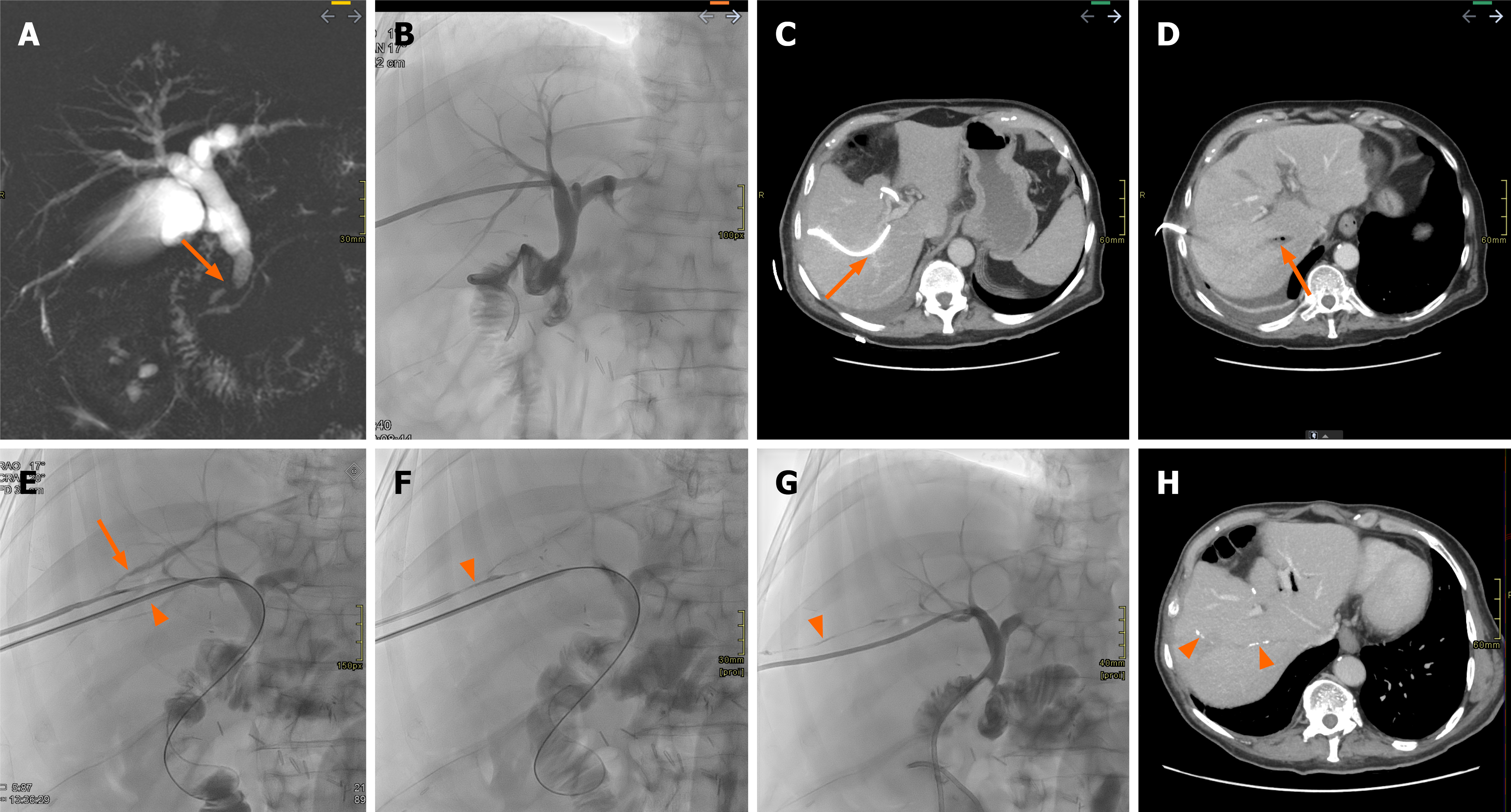
Figure 6 A 73-year-oid patient with cholangiocarcinoma and bile leak.
A: Magnetic resonance cholangiopancreatography demonstrates a filling defect in the distal common bile duct (arrow), later diagnosed as cholangiocarcimona. The patient subsequently underwent surgical resection with biliodigestive anastomosis; B: Bile leakage was observed from the surgical drain, prompting percutaneous transhepatic cholangiography. In the days following the percutaneous transhepatic cholangiography, the patient developed sepsis; C: Computed tomography was performed and revealed a marked angulation of the percutaneous drain in close proximity to vascular structures (arrow); D: Along with thrombosis and the presence of gas bubbles in the right suprahepatic vein (arrow); E: A repeat interventional radiology procedure was carried out, which confirmed the presence of a biliovenous leak (arrow) and a new drain was placed in a different right bile duct (arrowhead); F: Subsequently, the biliovenous fistula tract was embolized using N-butyl cyanoacrylate glue (arrowhead); G: Finally, the previously positioned access route was removed, with continued embolization of the entire access tract (arrowhead); H: On follow-up computed tomography after removal of the biliary drain and resolution of the patient’s clinical condition, faint remnants of the embolic material are still visible (arrowheads).
- Citation: Acquafredda F, Vizzuso A, Grigoriadis S, Di Giovanni G, Renzulli M, Giampalma E, Paraskevopoulos I, Spiliopoulos S, Inchingolo R. Vascular complications in percutaneous biliary interventions. World J Radiol 2026; 18(8): 119018
- URL: https://www.wjgnet.com/1949-8470/full/v18/i8/119018.htm
- DOI: https://dx.doi.org/10.4329/wjr.119018