
















Copyright: ©Author(s) 2026.
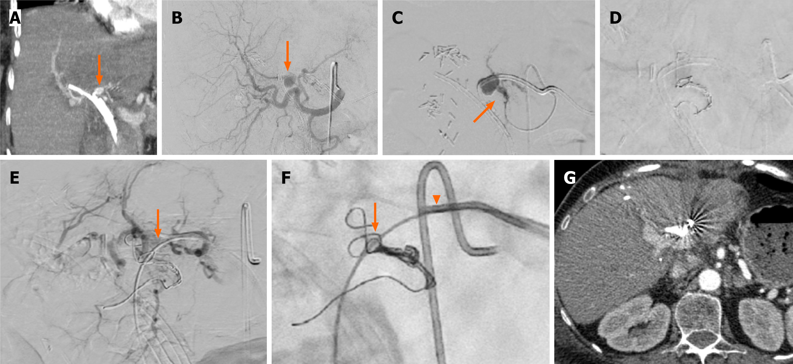
Figure 3 A 53-year-old female patient with colorectal liver hilar metastasis and right lobe endoscopic plastic stent underwent left lobe percutaneous transhepatic biliary drainage.
A: Computed tomography angiography revealed a pseudoaneurysm in proximity to the percutaneous biliary catheter (arrow); B: Selective digital subtraction angiography from the common hepatic artery confirmed the presence of the pseudoaneurysm arising from the left hepatic artery (arrow); C and D: The left hepatic artery was selectively catheterized with a 1.98Fr microcatheter and proximately embolized with pushable microcoils; as the back to front door” technique was not feasible (microcatheter was not advancing distal to the lesion) the catheter was removed over a 0.035’ guide wire (arrow), E and F: An 8Fr sheath was placed and using a 5Fr angled catheter (arrow) and the same microcatheter via the 5Fr catheter (arrowhead), the sack of the pseudoaneurysm was embolized using pushable microcoils (arrow). Note that during removal of the biliary catheter arterial blood was noted, confirming the necessity of further embolization from the biliary access; G: Check computed tomography angiography at 7 days, prior to discharge, confirmed the occlusion of the pseudoaneurysm. The patient was discharged asymptomatic.
- Citation: Acquafredda F, Vizzuso A, Grigoriadis S, Di Giovanni G, Renzulli M, Giampalma E, Paraskevopoulos I, Spiliopoulos S, Inchingolo R. Vascular complications in percutaneous biliary interventions. World J Radiol 2026; 18(8): 119018
- URL: https://www.wjgnet.com/1949-8470/full/v18/i8/119018.htm
- DOI: https://dx.doi.org/10.4329/wjr.119018