
















Copyright: ©Author(s) 2026.
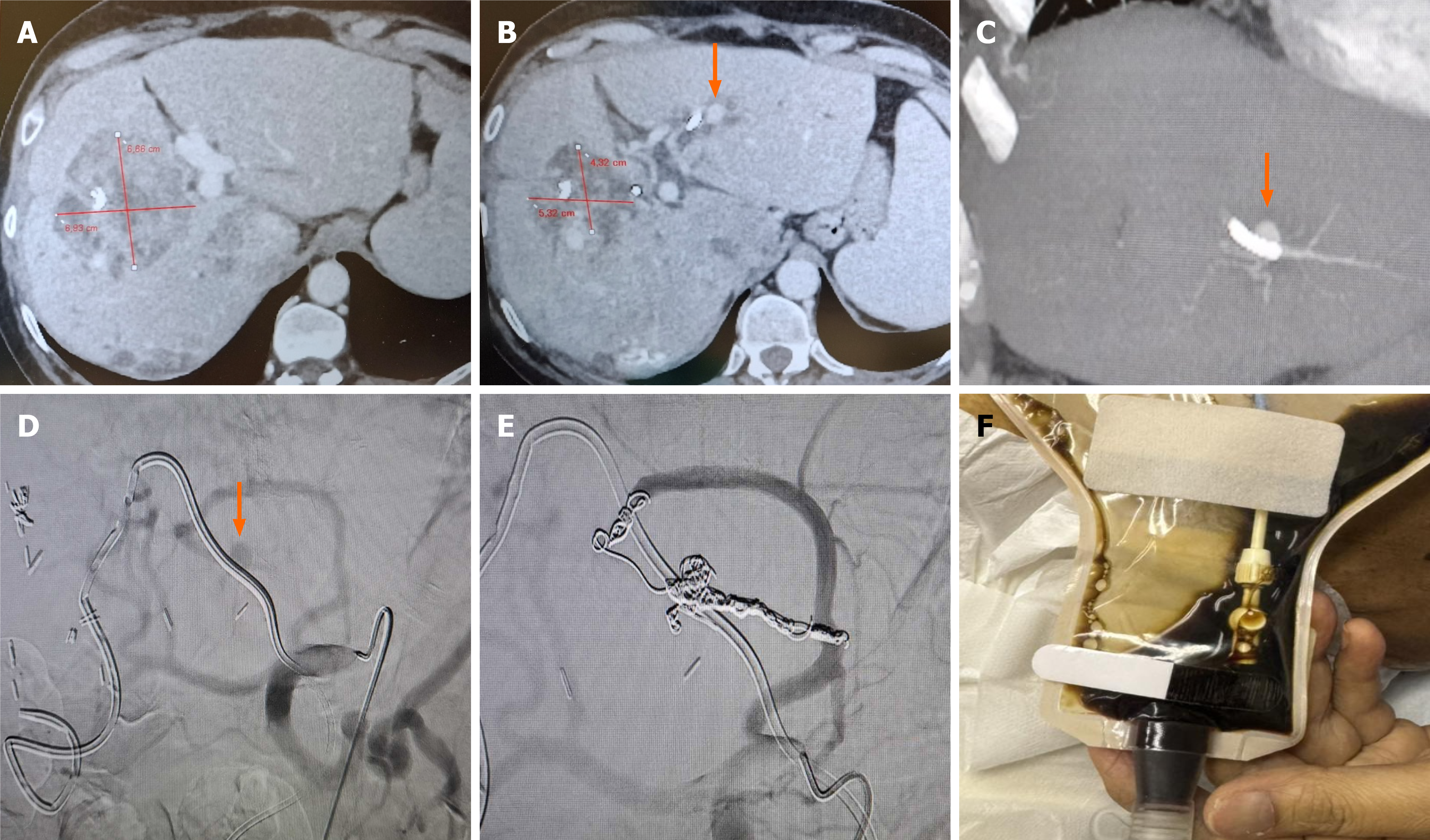
Figure 2 A 59-year-old female patient with obstructive jaundice due to three colorectal liver metastasis (max diameter of largest lesion 7 cm) underwent biliary drainage as to enable radioembolization therapy.
A: Access was obtained from the left lobe due to extensive right lobe metastatic disease. Selective Internal Radiation Therapy was successfully performed. However, 10 weeks after Selective Internal Radiation Therapy the patient reported blood within the percutaneous transhepatic biliary drainage collective bag. Following urgent admission hemobilia was diagnosed (right quadrant pain and melena); B and C: Computed tomography angiography depicted a pseudoaneurysm in proximity to the percutaneous biliary catheter (arrows), as well as good response to the radioembolization (treated lesions with significantly decreased perfusion and size); D and E: Selective digital subtraction angiography of the celiac axis verified the pseudoaneurysm which was arising from a left hepatic artery branch (arrow). Selective “back to front door” embolization was using a 1.98Fr microcatheter and detachable microcoils. F: At 48 hours bile in the collecting bag was clear of blood, blood tests normalized and the patient was discharged asymptomatic.
- Citation: Acquafredda F, Vizzuso A, Grigoriadis S, Di Giovanni G, Renzulli M, Giampalma E, Paraskevopoulos I, Spiliopoulos S, Inchingolo R. Vascular complications in percutaneous biliary interventions. World J Radiol 2026; 18(8): 119018
- URL: https://www.wjgnet.com/1949-8470/full/v18/i8/119018.htm
- DOI: https://dx.doi.org/10.4329/wjr.119018