
















Copyright: ©Author(s) 2026.
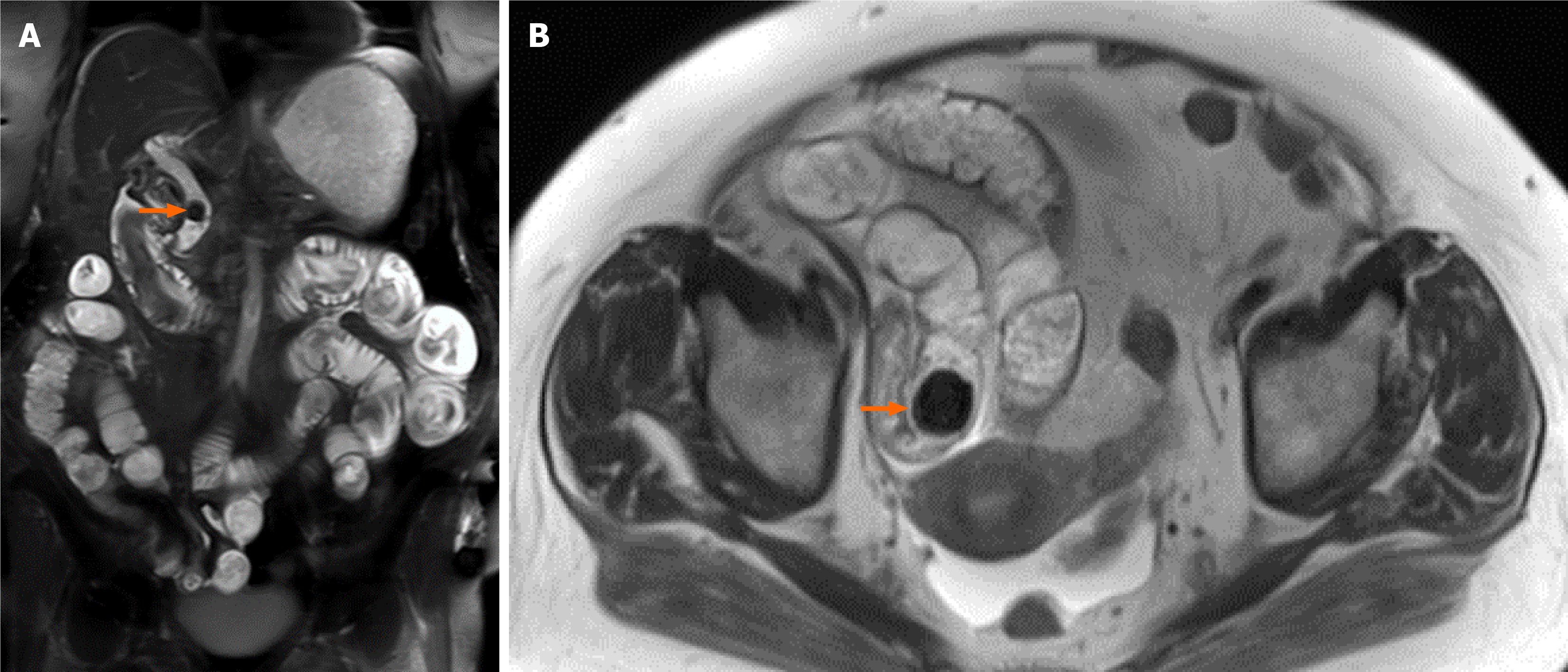
Figure 8 Gallstone ileus on magnetic resonance imaging.
A: Coronal T1 fat suppressed magnetic resonance imaging of the abdomen demonstrating a common bile duct stone (orange arrow) and diffusely distended small bowel loops; B: Axial T2 image showing an additional stone in the distal ileal loop causing proximal obstruction (orange arrow). These findings are in keeping with gallstone ileus.
- Citation: Gonzalez Baerga CI, Menendez Santos M, Ortiz Cordero RG, Guevara Tirado OA, Virarkar M, Gopireddy DR. Role of magnetic resonance in bowel pathologies: Beyond inflammatory bowel disease. World J Radiol 2026; 18(7): 122165
- URL: https://www.wjgnet.com/1949-8470/full/v18/i7/122165.htm
- DOI: https://dx.doi.org/10.4329/wjr.122165