
















Copyright: ©Author(s) 2026.
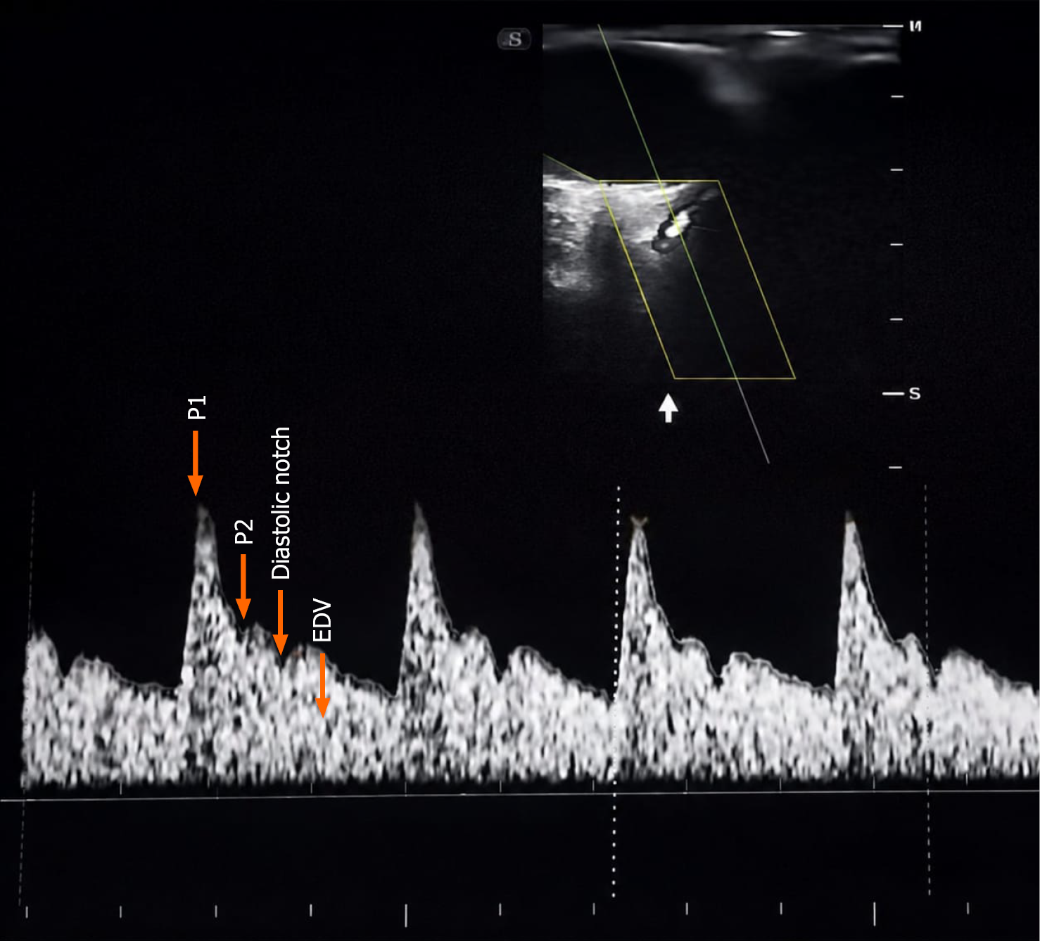
Figure 1
Ophthalmic artery Doppler image demonstrating the first systolic peak velocity (P1), second systolic peak velocity (P2), diastolic notch, and end-diastolic velocity (EDV).
- Citation: Dhiman R, Nayyar N, Takkar P, Sood D, Shukla A. Predictive value of second-trimester ophthalmic artery Doppler for identifying women at risk for preeclampsia. World J Radiol 2026; 18(7): 120995
- URL: https://www.wjgnet.com/1949-8470/full/v18/i7/120995.htm
- DOI: https://dx.doi.org/10.4329/wjr.120995