
















Copyright: ©Author(s) 2026.
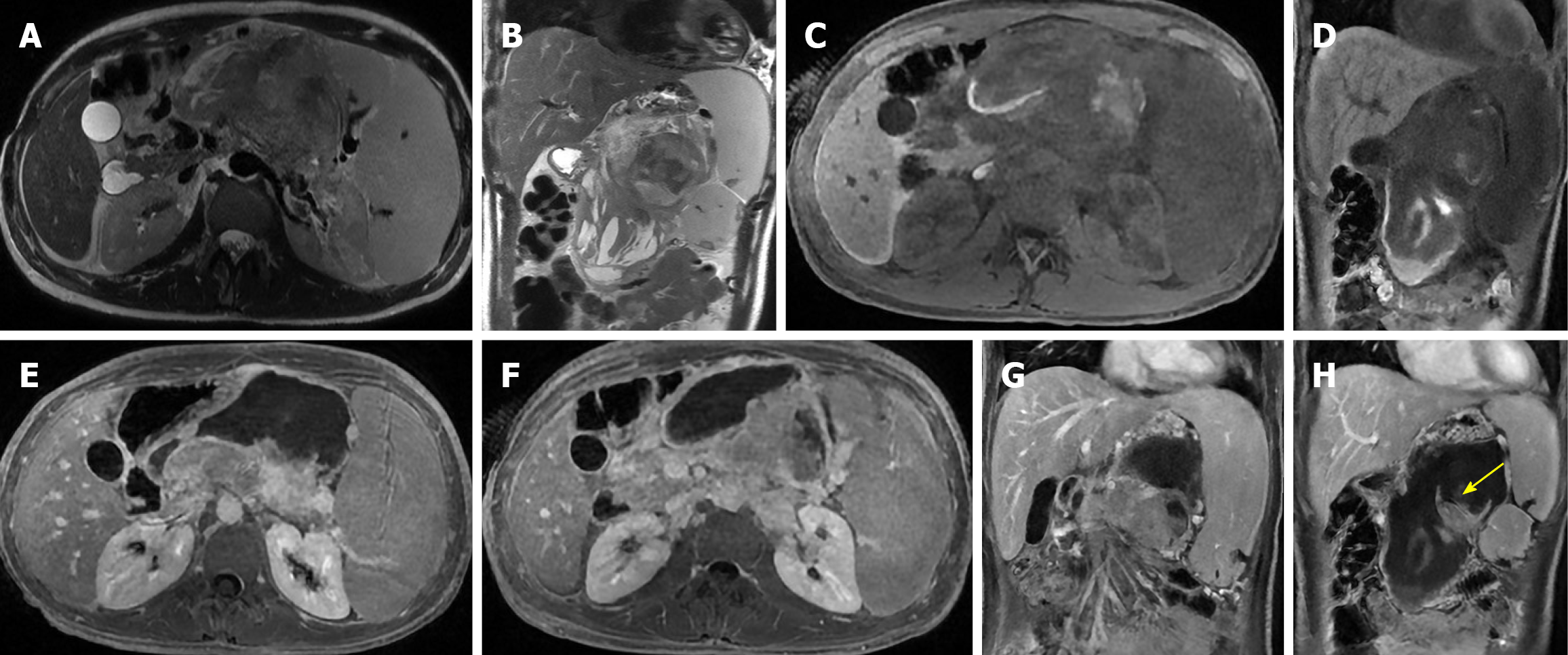
Figure 19 Large solid pseudopapillary neoplasm with spontaneous rupture and hemoperitoneum.
A and B: Axial and coronal T2-weighted images show a large heterogeneous pancreatic mass with mixed solid and cystic components and extensive intralesional hemorrhage; C and D: Axial and coronal T1-weighted images demonstrate intrinsic high signal intensity consistent with blood products; E and F: Axial post-contrast T1-weighted images show heterogeneous enhancement of the viable solid portions with non-enhancing hemorrhagic areas; G and H: Coronal post-contrast T1-weighted images confirm capsular disruption (arrow) with adjacent fluid collections consistent with hemoperitoneum. In a 16-year-old boy, these findings are consistent with spontaneous rupture of solid pseudopapillary neoplasm complicated by intra-tumoral hemorrhage and hemoperitoneum.
- Citation: Boraschi P, Cervelli R, Donati F. Common cystic pancreatic neoplasms: A comprehensive magnetic resonance imaging-based review of typical and atypical imaging features. World J Radiol 2026; 18(7): 120526
- URL: https://www.wjgnet.com/1949-8470/full/v18/i7/120526.htm
- DOI: https://dx.doi.org/10.4329/wjr.120526