
















Copyright: ©Author(s) 2026.
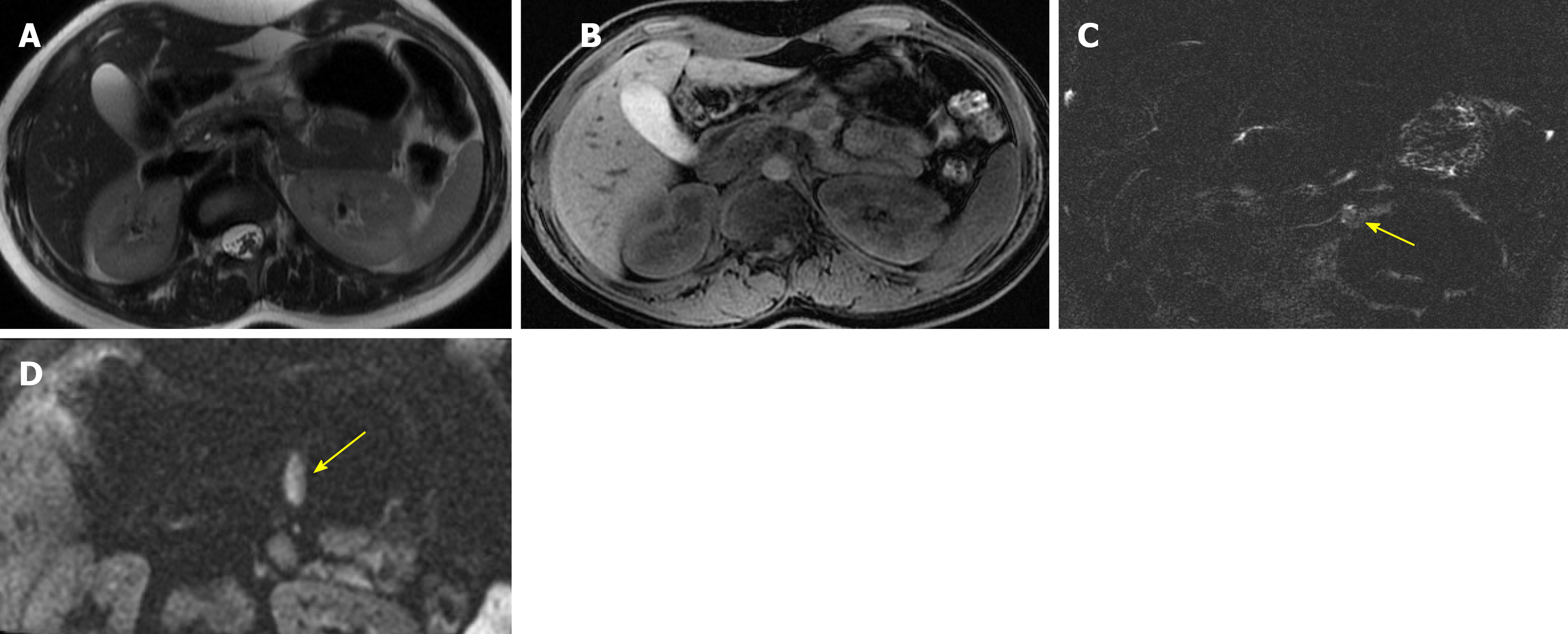
Figure 17 Atypical small solid variant of solid pseudopapillary neoplasm.
A: Axial T2-weighted image shows a small, well-defined solid lesion with mildly hyperintense signal in the pancreatic body; B: Axial T1-weighted image demonstrates low signal intensity; C: Thin-slab magnetic resonance cholangiopancreatography shows only poor hyperintensity (arrow), reflecting the predominantly solid nature of the lesion; D: Diffusion weighted imaging (b = 1000 second/mm2) demonstrates high hyperintensity on high b values (arrow). In a 34-year-old woman, this homogeneous solid appearance represents an atypical presentation of solid pseudopapillary neoplasm and may mimic a pancreatic neuroendocrine tumor.
- Citation: Boraschi P, Cervelli R, Donati F. Common cystic pancreatic neoplasms: A comprehensive magnetic resonance imaging-based review of typical and atypical imaging features. World J Radiol 2026; 18(7): 120526
- URL: https://www.wjgnet.com/1949-8470/full/v18/i7/120526.htm
- DOI: https://dx.doi.org/10.4329/wjr.120526