
















Copyright: ©Author(s) 2026.
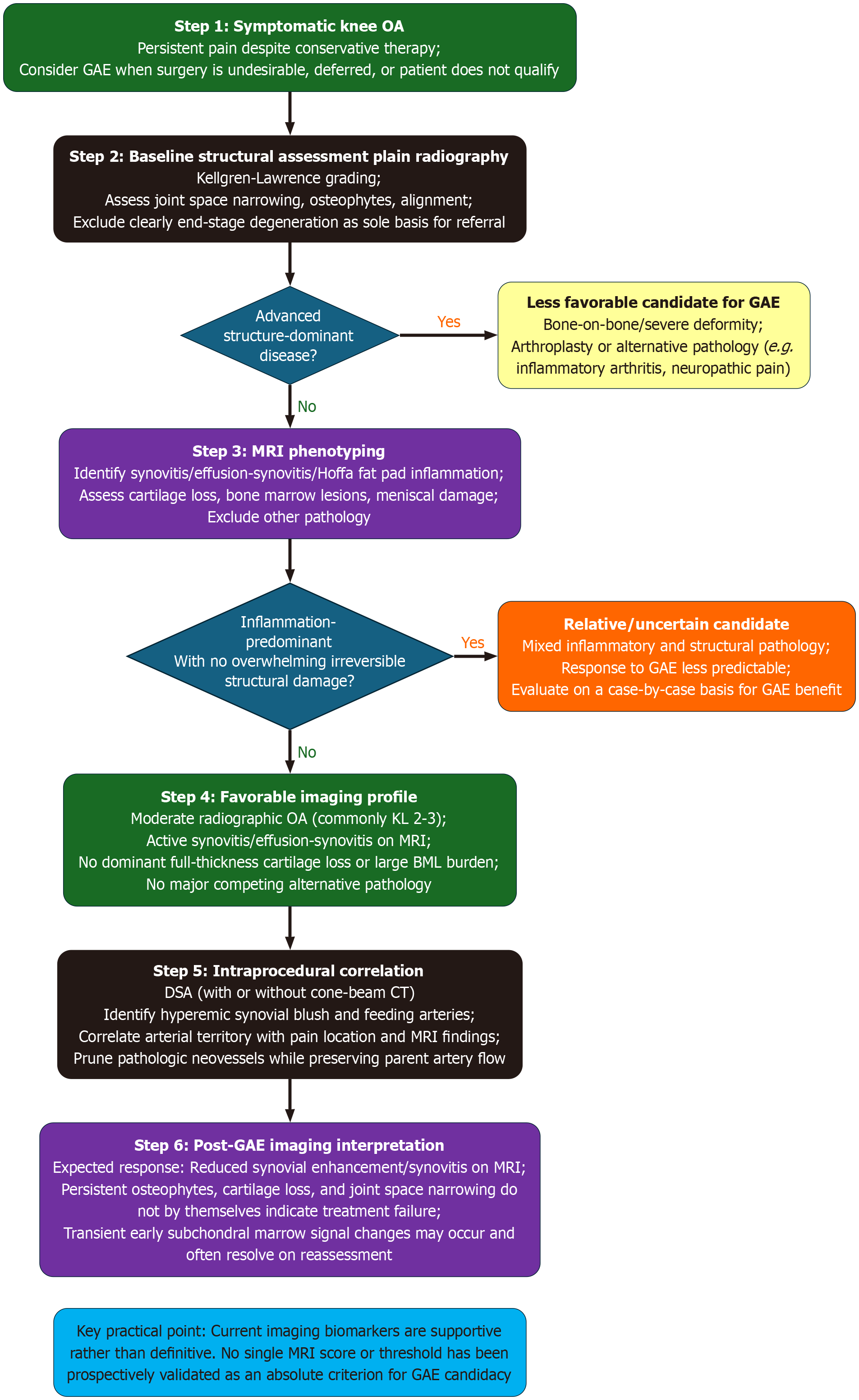
Figure 4 Proposed imaging-based algorithm for genicular artery embolization candidate selection and workflow in knee osteoarthritis.
Green boxes indicate start/proceed to next step, dark blue boxes are decision boxes, purple boxes are imaging boxes, yellow box is contraindication, and orange box is uncertainty/reevaluate. Plain radiography establishes structural severity, whereas magnetic resonance imaging (MRI) helps distinguish inflammation-predominant from structure-predominant disease. Favorable candidates generally show active synovitis-related MRI findings without overwhelming irreversible structural damage. Digital subtraction angiography, with or without cone-beam computed tomography, is then used to localize hyperemic synovial blush and confirm target vessels. Post-genicular artery embolization imaging response is expected to be predominantly inflammatory rather than structural. GAE: Genicular artery embolization; MRI: Magnetic resonance imaging; DSA: Digital subtraction angiography; CT: Computed tomography; OA: Osteoarthritis; KL: Kellgren-Lawrence.
- Citation: Singh R, Makary MS. Multimodality imaging considerations for genicular artery embolization in knee osteoarthritis. World J Radiol 2026; 18(6): 120315
- URL: https://www.wjgnet.com/1949-8470/full/v18/i6/120315.htm
- DOI: https://dx.doi.org/10.4329/wjr.120315