
















Copyright: ©Author(s) 2026.
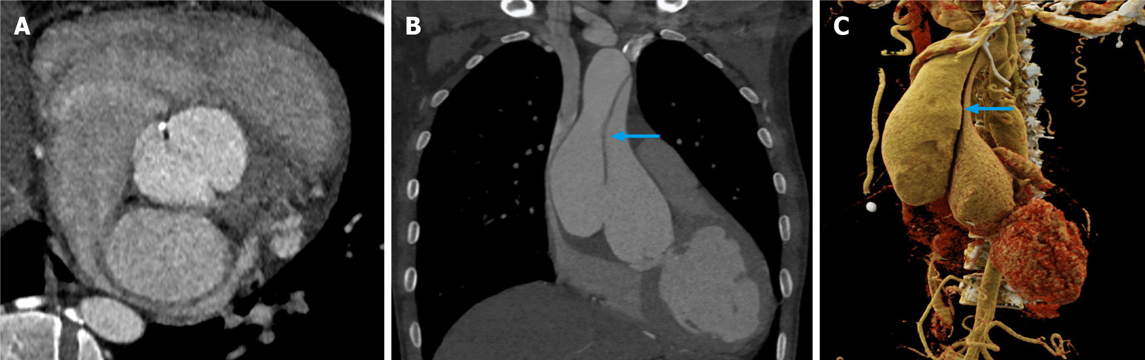
Figure 2 Bicuspid aortic valve with type A aortic dissection.
A: True axial view demonstrating type 0 bicuspid aortic valve; B and C: Coronal (B) and volume rendered image (C) depicting dilated ascending aorta with type A aortic dissection (shown by blue arrows). Images acquired during prospective elec
- Citation: Thanihaivel E S, Sharma A, Verma M, Vijayvergiya R, Singhal M. Evaluation of bicuspid aortic valve-associated aortopathic changes using third-generation dual-source computed tomography. World J Radiol 2026; 18(6): 120056
- URL: https://www.wjgnet.com/1949-8470/full/v18/i6/120056.htm
- DOI: https://dx.doi.org/10.4329/wjr.120056