
















Copyright: ©Author(s) 2026.
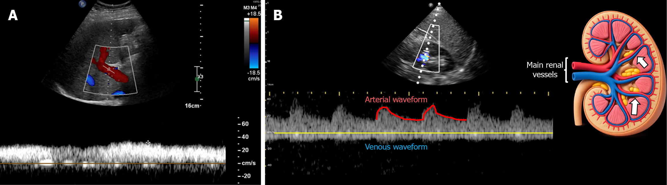
Figure 3 Doppler waveform.
A: Normal portal vein Doppler waveform demonstrating near-continuous flow with mild undulations above the baseline; B: Normal intrarenal venous Doppler waveform demonstrating continuous flow below the baseline with a concurrent arterial waveform above. The Doppler sample gate is positioned at the interlobar vessels, indicated by white arrows in the accompanying illustration.
- Citation: Shah C, Shah D, Koratala A. Venous excess doppler ultrasound: What radiologists should know in 2026. World J Radiol 2026; 18(6): 119110
- URL: https://www.wjgnet.com/1949-8470/full/v18/i6/119110.htm
- DOI: https://dx.doi.org/10.4329/wjr.119110