
















Copyright: ©Author(s) 2026.
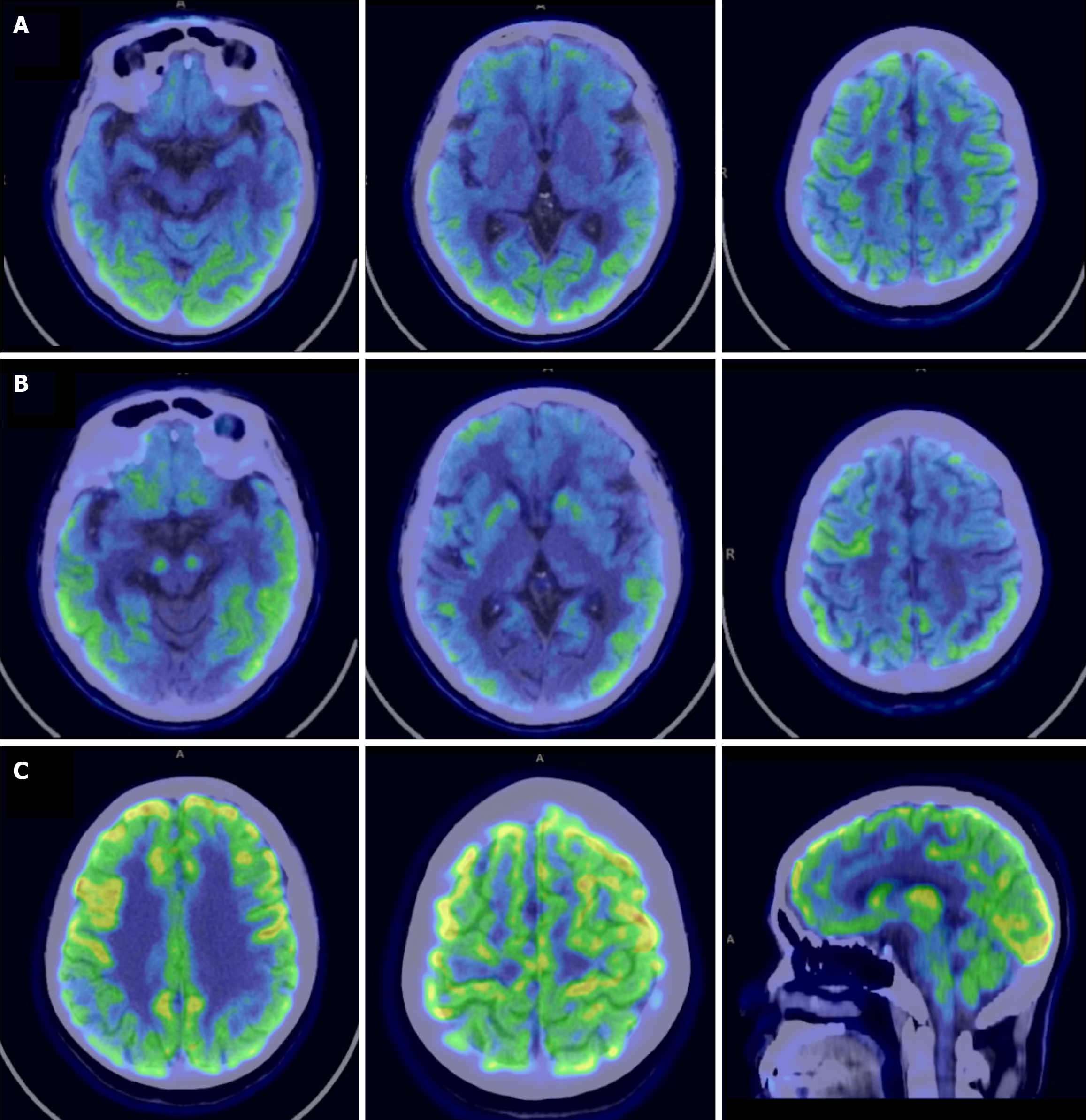
Figure 4 Positron emission topography scans of the proband.
A: 18F-florbetapir (AV-45) positron emission topography imaging showed extensive cerebral cortex and cerebellar amyloid beta deposition; B: 18F-flortaucipir (AV-1451) PET showed abnormal diffuse tau deposition throughout the bilateral frontal, temporal, parietal, and occipital lobes, and the cingulate gyrus; C: Fluorodeoxyglucose positron emission tomography-positron emission topography revealed diffuse hypometabolism in the right lateral temporal lobe, bilateral posterior cingulate gyri, bilateral precunei, right superior parietal lobule, and bilateral inferior parietal lobules (right lobes/Lobules more affected).
- Citation: Li J, Zhou FL, Chen JJ, Lin ZJ, Shi X, Zhang GG, Yang HJ, Dong MJ, Yi L, Hu J, Chen XH. Novel PSEN1 (Q223 L) mutation causes early-onset Alzheimer’s disease: A case report. World J Radiol 2026; 18(6): 116652
- URL: https://www.wjgnet.com/1949-8470/full/v18/i6/116652.htm
- DOI: https://dx.doi.org/10.4329/wjr.116652