















Copyright: ©Author(s) 2026.
World J Radiol. Apr 28, 2026; 18(4): 119851
Published online Apr 28, 2026. doi: 10.4329/wjr.v18.i4.119851
Published online Apr 28, 2026. doi: 10.4329/wjr.v18.i4.119851
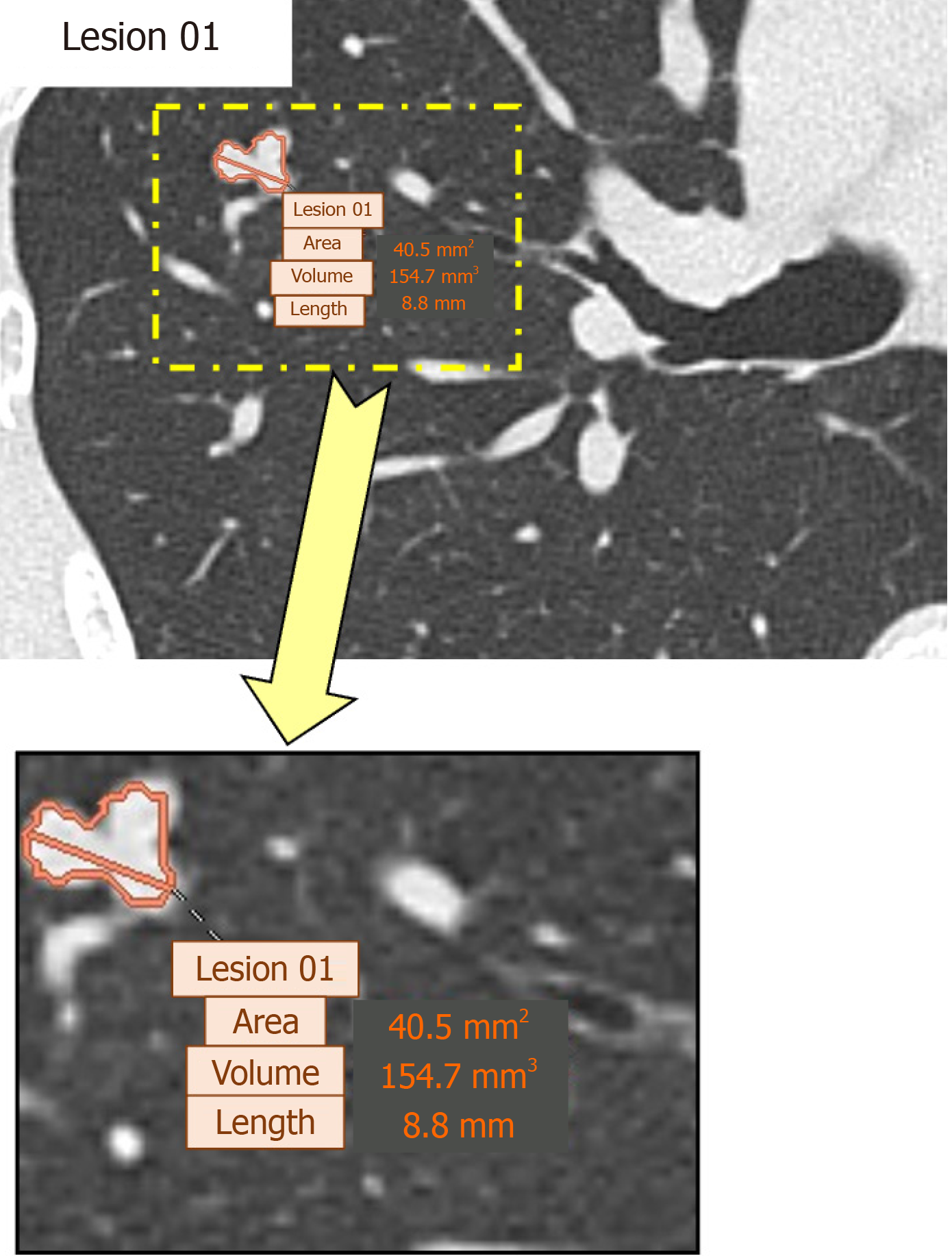
Figure 2 Detection and measurement of a pulmonary arteriovenous fistula using an artificial intelligence-based computer-aided detection system.
A representative example of a pulmonary arteriovenous fistula detected on chest computed tomography by the artificial intelligence-based computer-aided detection (CAD) system is shown. The CAD system automatically identified the pulmonary arteriovenous fistula and provided quantitative measurements, including maximum lesion length and lesion volume. The lower panel shows a magnified view of the detected lesion, demonstrating the quantitative parameters generated by the CAD system.
- Citation: Azama K, Tsuchiya N, Toyosato S, Yonemoto K, Nishie A. Artificial intelligence-based lung nodule detection for pulmonary arteriovenous fistulas on chest computed tomography. World J Radiol 2026; 18(4): 119851
- URL: https://www.wjgnet.com/1949-8470/full/v18/i4/119851.htm
- DOI: https://dx.doi.org/10.4329/wjr.v18.i4.119851