
















Copyright: ©Author(s) 2026.
World J Radiol. Apr 28, 2026; 18(4): 119319
Published online Apr 28, 2026. doi: 10.4329/wjr.v18.i4.119319
Published online Apr 28, 2026. doi: 10.4329/wjr.v18.i4.119319
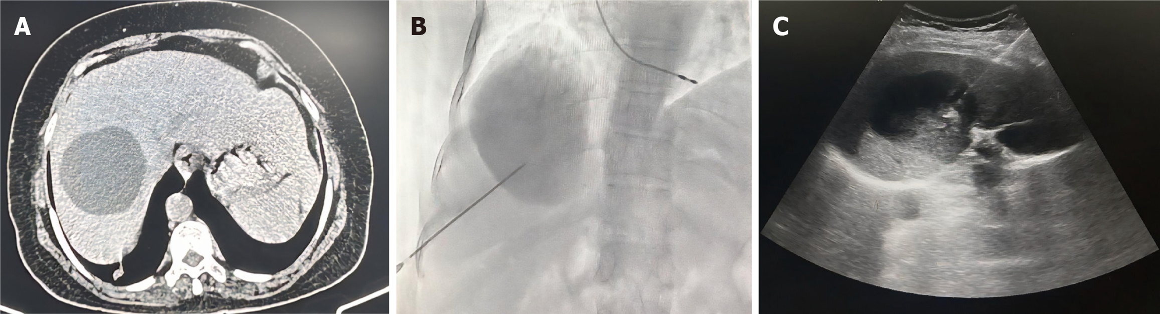
Figure 8 Örmeci technique.
A: Axial computed tomography image shows a hydatid cyst in the right lobe of the liver; B: Fluoroscopic spot image during Örmeci procedure: Contrast is injected through the puncture needle, which fills the cyst cavity with no opacification of biliary radicles, ruling out cysto-biliary communication; C: Ultrasound image depicting puncture needle in situ within the hydatid cyst.
- Citation: Agarwal D, Gadwal SK, Aswani Y, Das CJ. Role of interventional radiology in the management of hepatic hydatid disease. World J Radiol 2026; 18(4): 119319
- URL: https://www.wjgnet.com/1949-8470/full/v18/i4/119319.htm
- DOI: https://dx.doi.org/10.4329/wjr.v18.i4.119319