
















Copyright: ©Author(s) 2026.
World J Radiol. Apr 28, 2026; 18(4): 119319
Published online Apr 28, 2026. doi: 10.4329/wjr.v18.i4.119319
Published online Apr 28, 2026. doi: 10.4329/wjr.v18.i4.119319
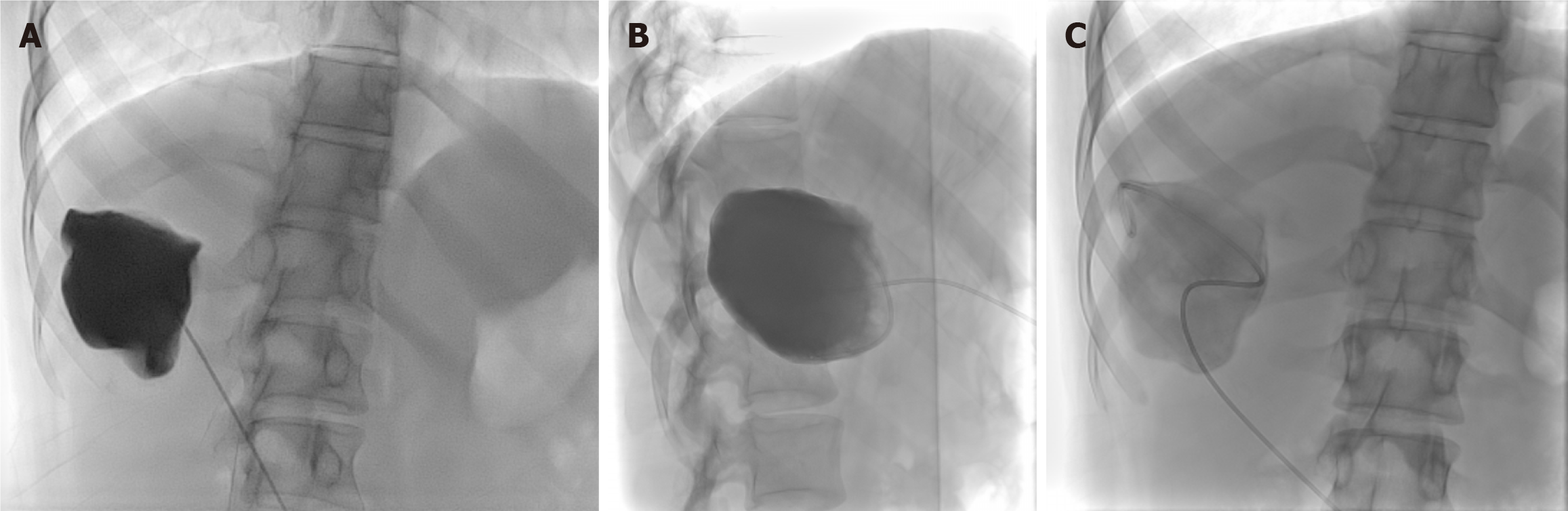
Figure 6 Puncture-aspiration-injection-reaspiration procedure.
A-C: Frontal fluoroscopic spot image (A) demonstrates intracystic injection of contrast, with no evidence of cysto-biliary communication. Lateral fluoroscopic spot image (B) after aspiration of the previously injected contrast from the hydatid cyst. Frontal fluoroscopic spot image (C) after instillation of scolicidal agent into the cyst cavity.
- Citation: Agarwal D, Gadwal SK, Aswani Y, Das CJ. Role of interventional radiology in the management of hepatic hydatid disease. World J Radiol 2026; 18(4): 119319
- URL: https://www.wjgnet.com/1949-8470/full/v18/i4/119319.htm
- DOI: https://dx.doi.org/10.4329/wjr.v18.i4.119319