
















Copyright: ©Author(s) 2026.
World J Radiol. Apr 28, 2026; 18(4): 119319
Published online Apr 28, 2026. doi: 10.4329/wjr.v18.i4.119319
Published online Apr 28, 2026. doi: 10.4329/wjr.v18.i4.119319
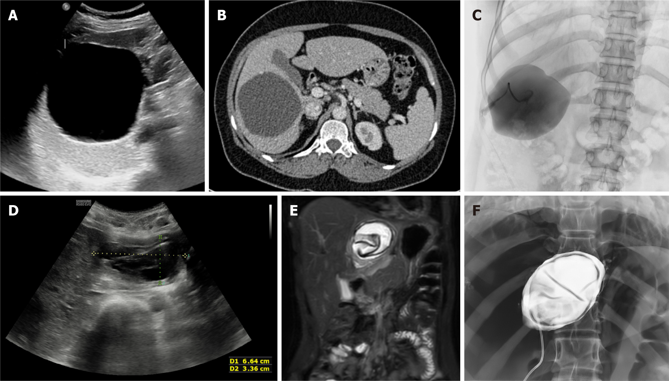
Figure 5 Puncture-aspiration-injection-reaspiration procedure.
A-C: Ultrasound (A) and axial computed tomography image (B) depict a cystic echinococcus 1 hydatid cyst in the right lobe of the liver. Fluoroscopic spot image (C) shows opacification of the cyst cavity following contrast injection, confirming correct intracystic placement with no evidence of cysto-biliary communication; D-F: Axial ultrasound (D) and coronal T2-weighted magnetic resonance imaging image (E) in a different patient depicts a cystic echinococcus 3a hydatid cyst in the left lobe of the liver. Fluoroscopic spot image (F) obtained during the puncture-aspiration-injection-reaspiration procedure shows opacification of the cyst cavity, with the floating membranes visualized as curvilinear filling defects, with no evidence of cysto-biliary communication.
- Citation: Agarwal D, Gadwal SK, Aswani Y, Das CJ. Role of interventional radiology in the management of hepatic hydatid disease. World J Radiol 2026; 18(4): 119319
- URL: https://www.wjgnet.com/1949-8470/full/v18/i4/119319.htm
- DOI: https://dx.doi.org/10.4329/wjr.v18.i4.119319