
















Copyright: ©Author(s) 2026.
World J Radiol. Mar 28, 2026; 18(3): 118143
Published online Mar 28, 2026. doi: 10.4329/wjr.v18.i3.118143
Published online Mar 28, 2026. doi: 10.4329/wjr.v18.i3.118143
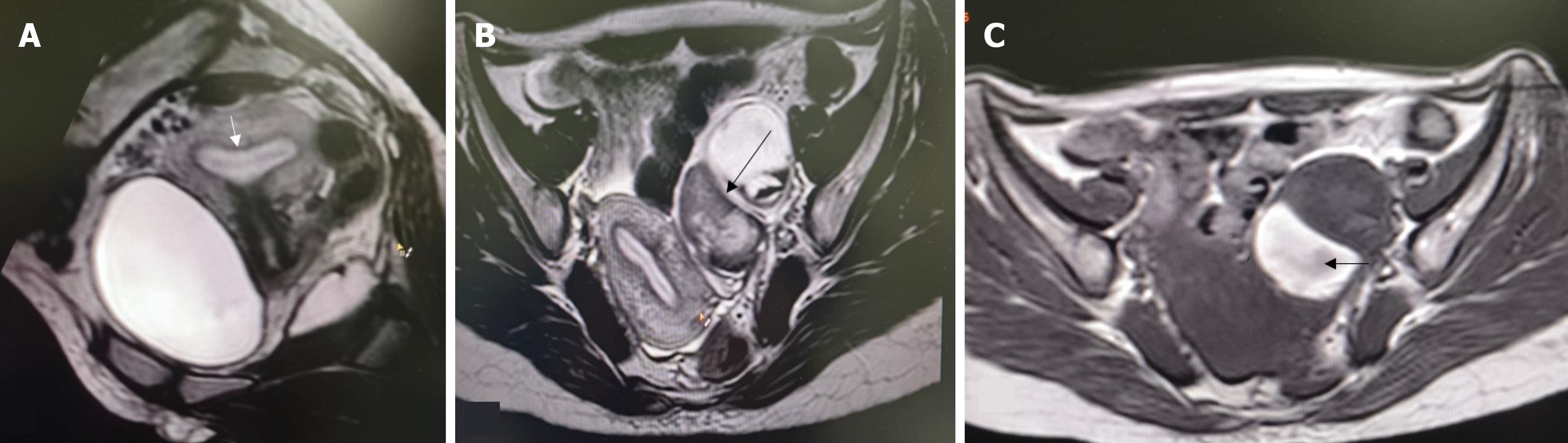
Figure 3 Arcuate uterus.
A: 3d T2W sagittal oblique image showing arcuate morphology of uterus (white arrow); B and C: Axial T2W image (B) and axial T1W image (C) showing left tubo-ovarian endometriotic complex with T1 hyperintense/T2 hypointense blood products (black arrow).
- Citation: Kumar I, Kumari S, Ojha R, Kumari R, Kushwaha K, Singh PK, Verma A. Characterization of endometriosis in patients with Mullerian anomalies using the Enzian classification: A retrospective cohort study. World J Radiol 2026; 18(3): 118143
- URL: https://www.wjgnet.com/1949-8470/full/v18/i3/118143.htm
- DOI: https://dx.doi.org/10.4329/wjr.v18.i3.118143