
















Copyright: ©Author(s) 2026.
World J Radiol. Mar 28, 2026; 18(3): 118143
Published online Mar 28, 2026. doi: 10.4329/wjr.v18.i3.118143
Published online Mar 28, 2026. doi: 10.4329/wjr.v18.i3.118143
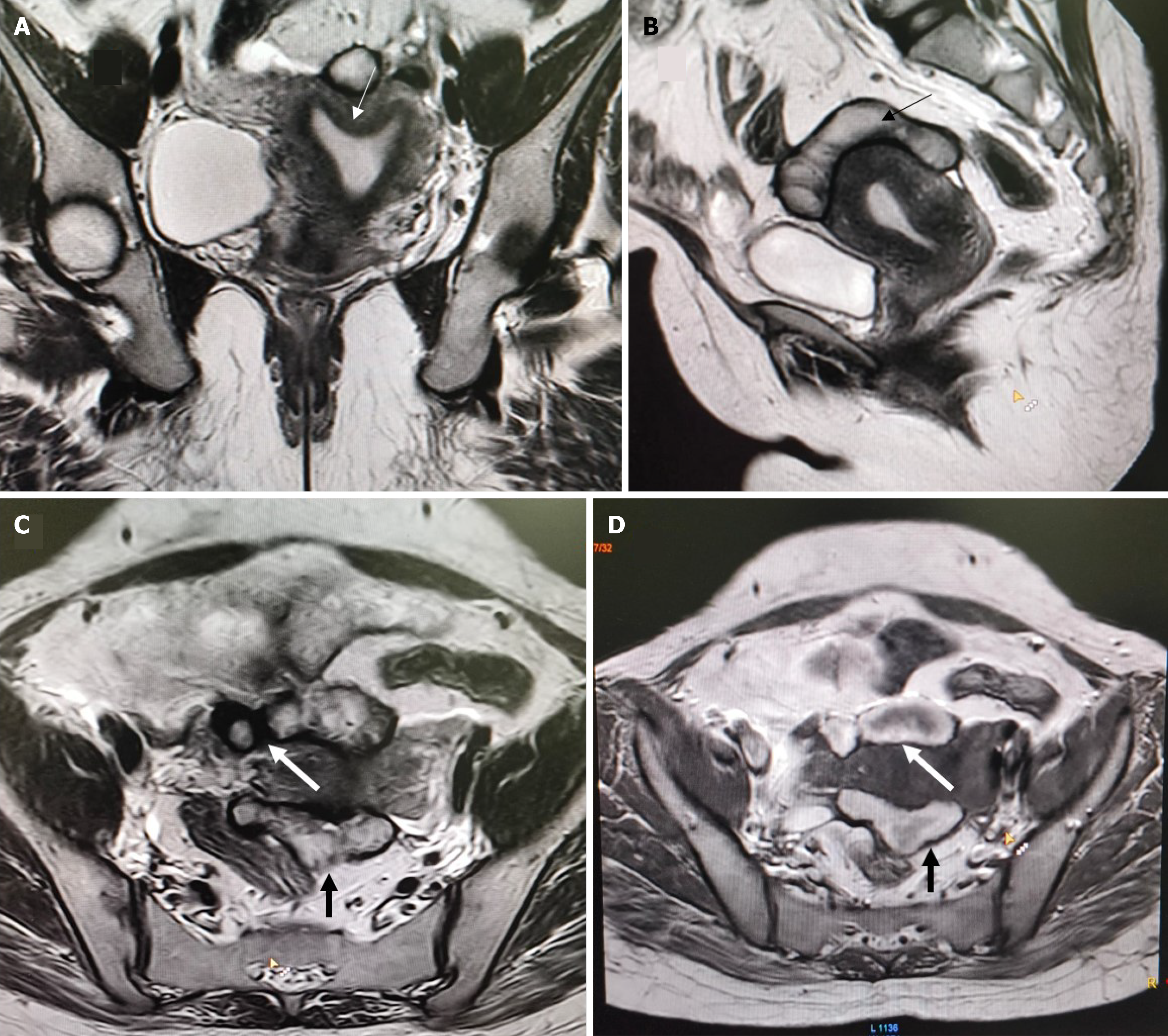
Figure 1 Arcuate uterus.
A: Coronal T2W image showing arcuate uterus morphology of internal indentation < 1 cm (arrow); B: Sagittal T2W image showing dilated left fallopian tube with hypointense rim and hematosalpinx (black arrow); C and D: Axial T2W (C) and axial T1W images (D) showing right (white thick arrows) and left (black thick arrow) endometriotic complex cysts coursing along anterior and posterior margins of uterus respectively with T1/T2 hyperintense signal consistent with blood products.
- Citation: Kumar I, Kumari S, Ojha R, Kumari R, Kushwaha K, Singh PK, Verma A. Characterization of endometriosis in patients with Mullerian anomalies using the Enzian classification: A retrospective cohort study. World J Radiol 2026; 18(3): 118143
- URL: https://www.wjgnet.com/1949-8470/full/v18/i3/118143.htm
- DOI: https://dx.doi.org/10.4329/wjr.v18.i3.118143