
















Copyright: ©Author(s) 2026.
World J Radiol. Mar 28, 2026; 18(3): 118119
Published online Mar 28, 2026. doi: 10.4329/wjr.v18.i3.118119
Published online Mar 28, 2026. doi: 10.4329/wjr.v18.i3.118119
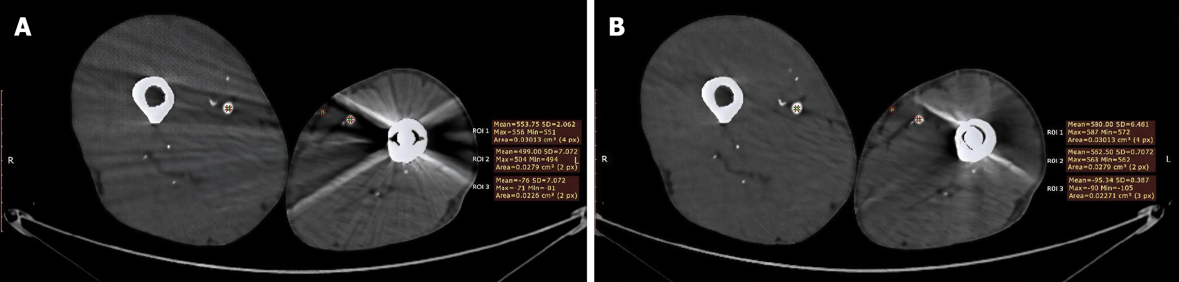
Figure 1 Improved femoral artery visualization after metal artifact reduction software processing.
A: Pre-metal artifact reduction (MAR), lower limbs axial computed tomography angiography without MAR software demonstrates streak and bloom artifacts originating from left femoral intra-medullary nail. Three regions of interest (ROI) were placed; ROI 1 in the contralateral (right) femoral artery shows mean Hounsfield unit (HU) of 553.75, SD = 2.062, ROI 2 in the ipsilateral (left) femoral artery demonstrates suboptimal contrast opacification, mean HU of 499.0, SD = 7.072. ROI 3 was placed in left subcutaneous fat with mean HU of -76, SD = 7.072; B: Post-MAR, same anatomic level post MAR software processing demonstrates marked artifact reduction with improved visualization of femoral artery. ROI 1 in the contralateral (right) femoral artery shows mean HU of 580.0, SD = 6.481, ROI 2 in the ipsilateral (left) femoral artery demonstrates suboptimal contrast opacification, mean HU of 562.5.0, SD = 7.072. ROI 3 was placed in left subcutaneous fat with mean HU of -95.3, SD = 8.387. ROI: Regions of interest.
- Citation: Mounir AM, Elmokadem AH, Saleh GAH, El-Morsy A, Abd El-Raouf GH. Role of metal artifact reduction software in computed tomography angiography of lower limb metallic prosthesis: A retrospective study. World J Radiol 2026; 18(3): 118119
- URL: https://www.wjgnet.com/1949-8470/full/v18/i3/118119.htm
- DOI: https://dx.doi.org/10.4329/wjr.v18.i3.118119