
















©Author(s) (or their employer(s)) 2026.
World J Radiol. Feb 28, 2026; 18(2): 116610
Published online Feb 28, 2026. doi: 10.4329/wjr.v18.i2.116610
Published online Feb 28, 2026. doi: 10.4329/wjr.v18.i2.116610
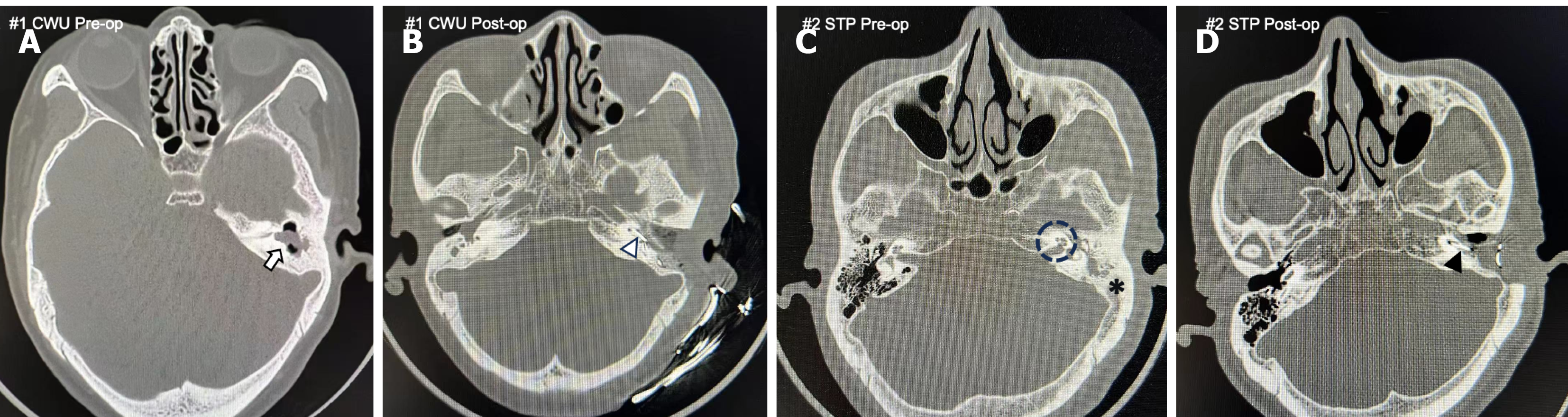
Figure 2 Representative preoperative high-resolution computed tomography dictates surgical strategy and correlates with postoperative outcomes.
A: Well-defined soft-tissue opacity (hollow arrow) confined to the epitympanum, with a well-aerated mastoid; B: Fully inserted cochlear implant electrode array (hollow arrowhead) with preserved ear canal anatomy after successful canal wall-up mastoidectomy; C: Complete opacification of the middle ear and mastoid (solid asterisk), with severe cochlear ossification (dotted circle) and posterior canal wall erosion; D: Obliterated mastoid cavity and partially inserted electrode array (solid arrowhead) following subtotal petrosectomy. CWU: Canal wall-up; Pre-op: Pre-operative; Post-op: Post-operative; STP: Subtotal petrosectomy.
- Citation: Zhao LJ, Fu Y, Zhang ZL. High-resolution computed tomography predicts optimal cochlear implantation strategy in patients with chronic otitis media. World J Radiol 2026; 18(2): 116610
- URL: https://www.wjgnet.com/1949-8470/full/v18/i2/116610.htm
- DOI: https://dx.doi.org/10.4329/wjr.v18.i2.116610