
















©Author(s) (or their employer(s)) 2026.
World J Radiol. Feb 28, 2026; 18(2): 116486
Published online Feb 28, 2026. doi: 10.4329/wjr.v18.i2.116486
Published online Feb 28, 2026. doi: 10.4329/wjr.v18.i2.116486
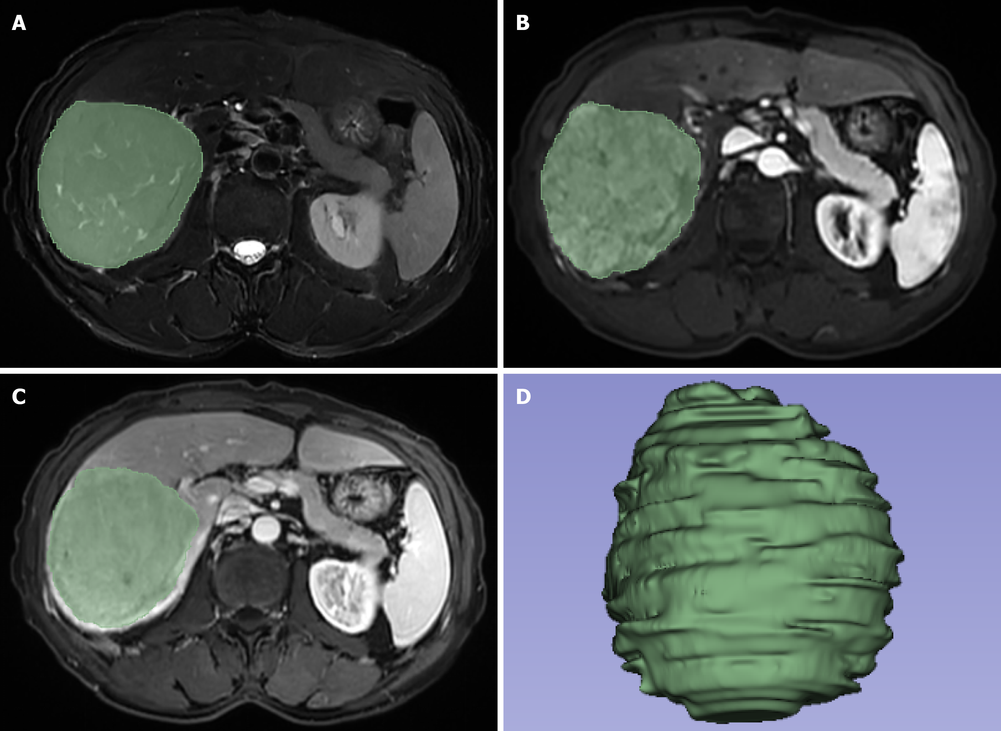
Figure 1 Region of interests along the edge of the lesion.
A: Fat-saturation T2-weighted imaging; B: Arterial phase image; C: Portal venous phase image; D: Three-dimensional image of the lesion.
- Citation: Mao Q, Zhang P, Zhou MT, Shi Y, Min XL, Xu H, Yang L, Zhang XM. Interpretable radiomics model based on magnetic resonance imaging to predict responses to transarterial chemoembolization for hepatocellular carcinoma. World J Radiol 2026; 18(2): 116486
- URL: https://www.wjgnet.com/1949-8470/full/v18/i2/116486.htm
- DOI: https://dx.doi.org/10.4329/wjr.v18.i2.116486