
















©Author(s) (or their employer(s)) 2026.
World J Radiol. Feb 28, 2026; 18(2): 115741
Published online Feb 28, 2026. doi: 10.4329/wjr.v18.i2.115741
Published online Feb 28, 2026. doi: 10.4329/wjr.v18.i2.115741
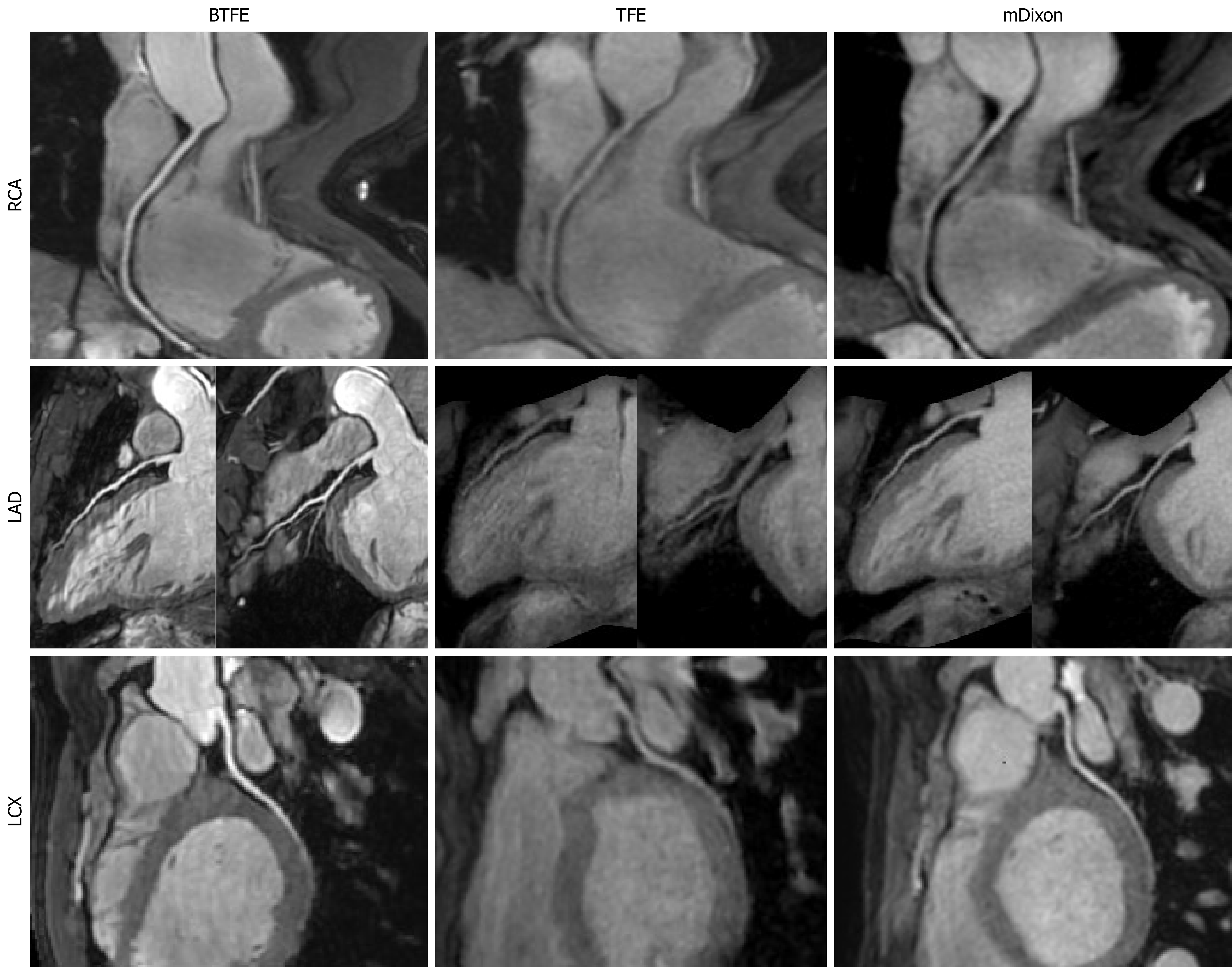
Figure 2 Non-contrast coronary magnetic resonance angiography images show the three major coronary arteries across the balanced turbo field echo, turbo field echo, and modified Dixon sequences.
The balanced turbo field echo (BTFE) sequence demonstrated superior image quality for all three arteries. In the right coronary artery, subjective image quality scores were 4 for BTFE, 2 for turbo field echo (TFE), and 3 for modified Dixon. The measured length of the left anterior descending artery was greater on BTFE images than on those acquired with TFE and modified Dixon. The lower contrast-to-noise ratio of the left circumflex artery on TFE likely explains its reduced measured length on that sequence. BTFE: Balanced turbo field echo; TFE: Turbo field echo; mDixon: Modified Dixon; LAD: Left anterior descending artery; LCX: Left circumflex artery; RCA: Right coronary artery.
- Citation: Wei CW, Zhang RZ, Xu Y, Liu JY, Zhang N, Xu L, Wen ZY. Non-contrast 3.0T coronary magnetic resonance angiography: Image quality comparison of coronal balanced-turbo-field-echo, axial turbo-field-echo, and modified-Dixon sequences. World J Radiol 2026; 18(2): 115741
- URL: https://www.wjgnet.com/1949-8470/full/v18/i2/115741.htm
- DOI: https://dx.doi.org/10.4329/wjr.v18.i2.115741