
















©Author(s) (or their employer(s)) 2026.
World J Radiol. Feb 28, 2026; 18(2): 115741
Published online Feb 28, 2026. doi: 10.4329/wjr.v18.i2.115741
Published online Feb 28, 2026. doi: 10.4329/wjr.v18.i2.115741
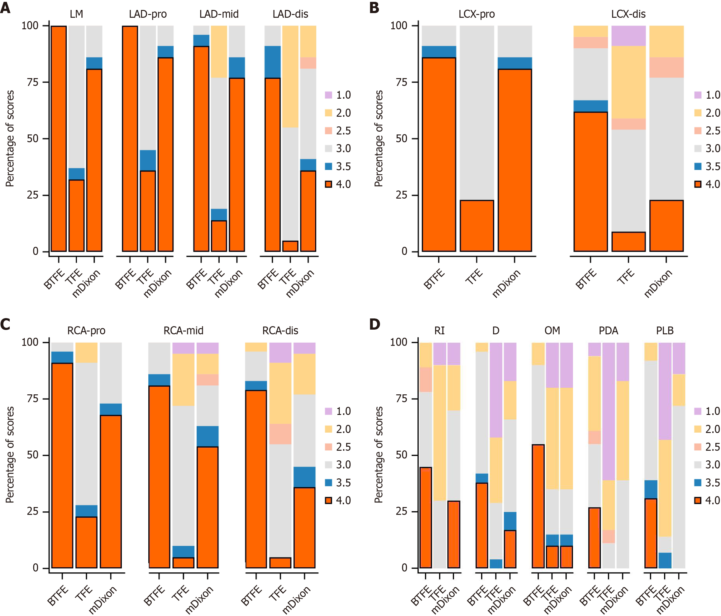
Figure 1 Stacked bar charts show image quality scores of three coronary magnetic resonance angiography sequences.
The stacked bar charts depict the distribution of subjective image quality scores (1-4) across 18 coronary segments for the three coronary magnetic resonance angiography sequences (balanced turbo field echo, turbo field echo, and modified Dixon). A: The stacked bar chart depict the distribution of subjective image quality scores (1-4) across left main coronary artery and the proximal, mid, and distal segments of the left anterior descending artery for the three coronary sequences; B: The stacked bar chart depict the distribution of subjective image quality scores (1-4) across the proximal and distal segments of the left circumflex artery for the three coronary sequences; C: The stacked bar chart depict the distribution of subjective image quality scores (1-4) across the proximal, mid, and distal segments of the right coronary artery for the three coronary sequences; D: The stacked bar chart depict the distribution of subjective image quality scores (1-4) across ramus intermediu, diagonal branch, obtuse marginal branch, posterior descending artery and posterolateral branch for the three coronary sequences. BTFE: Balanced turbo field echo; TFE: Turbo field echo; mDixon: Modified Dixon; LM: Left main artery; LAD-pro: Proximal left anterior descending artery; LAD-mid: Mid left anterior descending artery; LAD-dis: Distal left anterior descending artery; LCX-pro: Proximal left circumflex artery; LCX-dis: Distal left circumflex artery; RCA-pro: Proximal right coronary artery; RCA-mid: Mid right coronary artery; RCA-dis: Distal right coronary artery; RI: Ramus intermediu; D: The first and second diagonal branches (D1, D2); OM: The first and second obtuse marginal branches (OM1, OM2); PDA: Posterior descending arteries; PLB: Posterolateral branches.
- Citation: Wei CW, Zhang RZ, Xu Y, Liu JY, Zhang N, Xu L, Wen ZY. Non-contrast 3.0T coronary magnetic resonance angiography: Image quality comparison of coronal balanced-turbo-field-echo, axial turbo-field-echo, and modified-Dixon sequences. World J Radiol 2026; 18(2): 115741
- URL: https://www.wjgnet.com/1949-8470/full/v18/i2/115741.htm
- DOI: https://dx.doi.org/10.4329/wjr.v18.i2.115741