
















Copyright: ©Author(s) 2026.
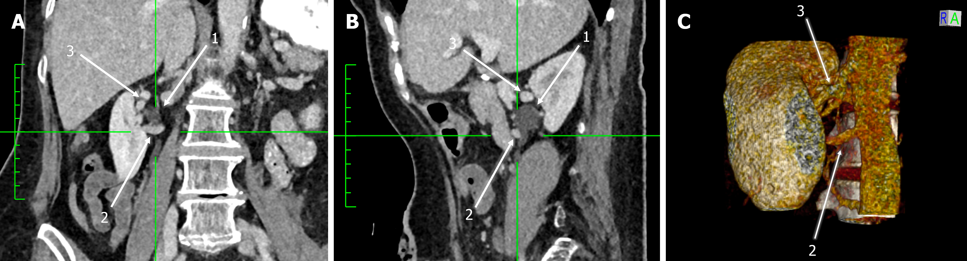
Figure 2 Computed tomography scan of 54-year-old male patient exhibiting bilateral anterior crossing by early bifurcated renal arteries at the level of ureteropelvic junction (on the left side) and 12.
5 mm distally on the ureter (on the right side). A: Coronal; B: Sagittal; C: Three-dimensional reconstruction. 1: Left ureter, 2: Left early bifurcated renal artery; 3: Right ureter.
- Citation: Paschopoulos I, Triantafyllou G, Gondorf D, Arkoudis NA, Velonakis G, Mariorakis C, Lyrtzis C, Gyftopoulos K, Piagkou M. Aberrant renal vessels crossing the ureter and the ureteropelvic junction: A retrospective computed tomography-based analysis of non-obstructed population. World J Radiol 2026; 18(10): 127348
- URL: https://www.wjgnet.com/1949-8470/full/v18/i10/127348.htm
- DOI: https://dx.doi.org/10.4329/wjr.127348