
















Copyright: ©Author(s) 2026.
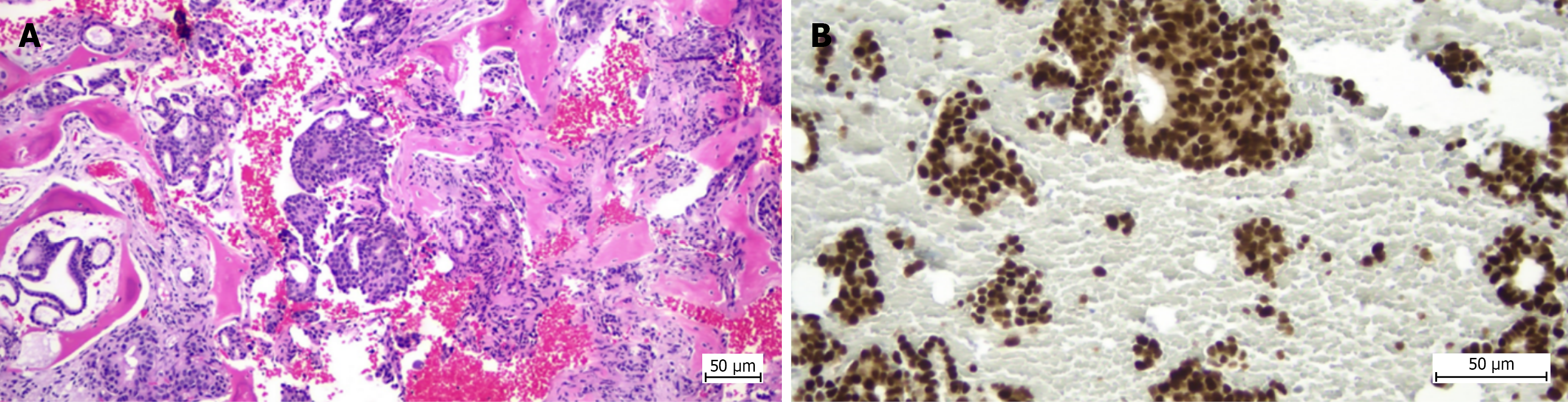
Figure 7 Histopathological and immunohistochemical appearance of the biopsy specimen in Case 2.
A: Fragments of trabecular bone tissue (black arrow) in which malignant neoplastic epithelial proliferation is identified, consisting of tubular, glandular and cribriform structures with rare solid structures (clusters, nests) and isolated cells, embedded in a desmoplastic stroma (hematoxylin and eosin staining, 100 × optical zoom); B: Estrogen receptor expression demonstrated by immunohistochemical staining, with intense and diffuse positivity in tumor cell nuclei (200 × optical zoom).
- Citation: Stuparu Adam AM, Osama A, Stere A, Cochior D, Pogărăşteanu ME, Moldovan CA, Tudorache IS, Adam R, Moldovan AD. Diagnostic performance of computed tomography-guided percutaneous bone biopsy in musculoskeletal oncology: A single-center cohort study. World J Radiol 2026; 18(10): 125901
- URL: https://www.wjgnet.com/1949-8470/full/v18/i10/125901.htm
- DOI: https://dx.doi.org/10.4329/wjr.125901