
















Copyright: ©Author(s) 2026.
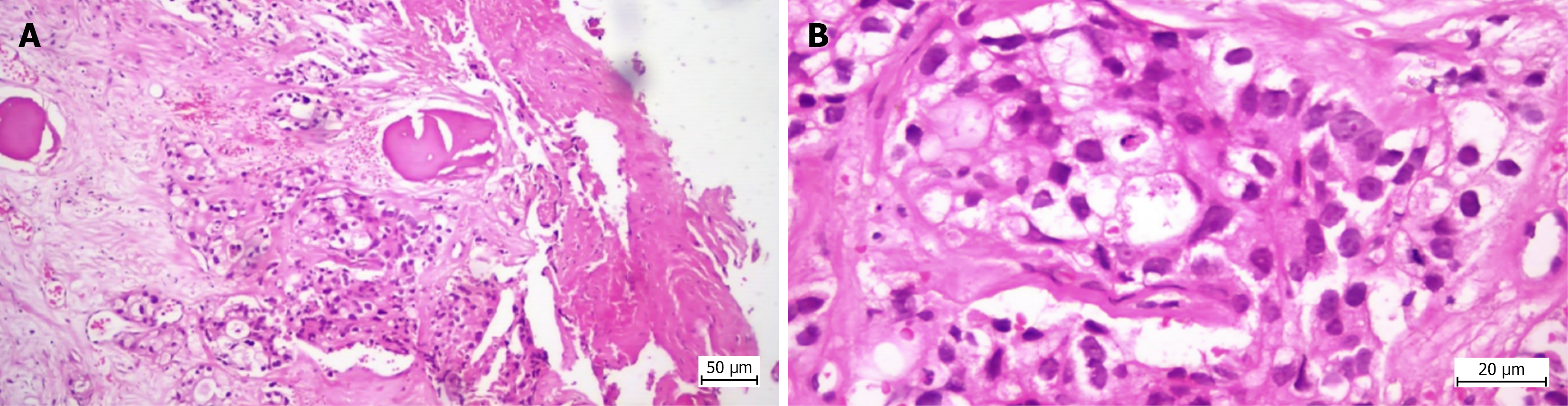
Figure 5 Histopathological appearance of the biopsy specimen in Case 1.
Bone tissue showing histological structure altered by malignant epithelial neoplastic proliferation with the histopathological appearance of poorly differentiated adenocarcinoma (G3) with glandular (distinct and cribriform) and solid development pattern. A: Hematoxylin and eosin staining, 100 × optical zoom; B: Hematoxylin and eosin staining, 400 × optical zoom.
- Citation: Stuparu Adam AM, Osama A, Stere A, Cochior D, Pogărăşteanu ME, Moldovan CA, Tudorache IS, Adam R, Moldovan AD. Diagnostic performance of computed tomography-guided percutaneous bone biopsy in musculoskeletal oncology: A single-center cohort study. World J Radiol 2026; 18(10): 125901
- URL: https://www.wjgnet.com/1949-8470/full/v18/i10/125901.htm
- DOI: https://dx.doi.org/10.4329/wjr.125901