
















©The Author(s) 2026.
World J Radiol. Jan 28, 2026; 18(1): 115504
Published online Jan 28, 2026. doi: 10.4329/wjr.v18.i1.115504
Published online Jan 28, 2026. doi: 10.4329/wjr.v18.i1.115504
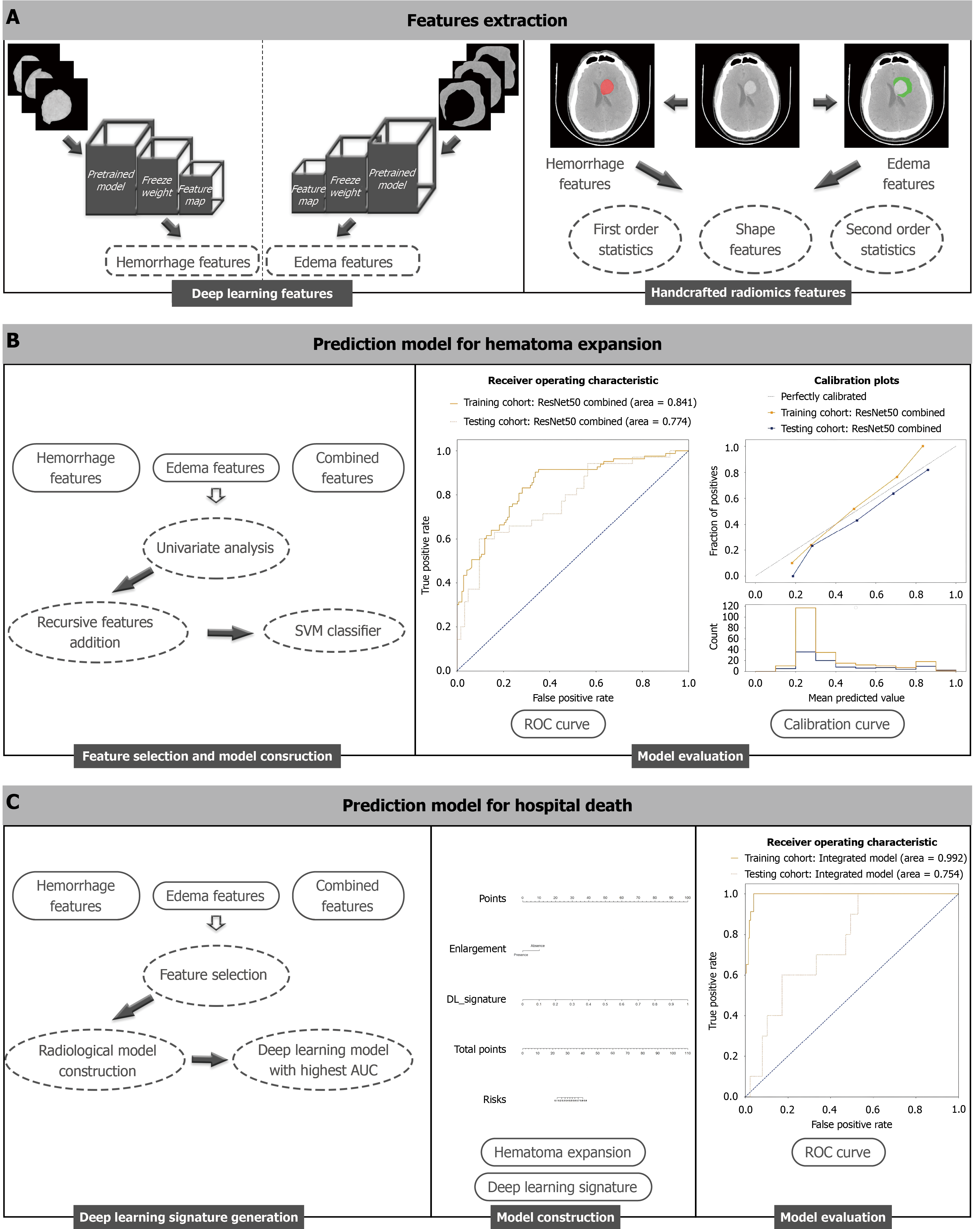
Figure 1 A general flowchart of data analysis.
A: Radiological features from hemorrhage and perihematomal edema non-contrast computed tomography images were extracted by the deep learning analysis and handcrafted radiomics analysis, respectively; B: The prediction models in identification of early enlargement of spontaneous intracerebral hemorrhage were approached via machine learning methods using radiological features; C: The prognostic model in prediction of hospital death took radiological features and the effect of hematoma expansion into account, and was visualized by nomogram. ROC: Receiver operating characteristic; SVM: Support vector machine.
- Citation: Yang YH, Li Y. Deep learning-based imaging model to predict early hematoma enlargement and hospital mortality in spontaneous intracerebral hemorrhage. World J Radiol 2026; 18(1): 115504
- URL: https://www.wjgnet.com/1949-8470/full/v18/i1/115504.htm
- DOI: https://dx.doi.org/10.4329/wjr.v18.i1.115504