
















©The Author(s) 2025.
World J Radiol. Sep 28, 2025; 17(9): 112983
Published online Sep 28, 2025. doi: 10.4329/wjr.v17.i9.112983
Published online Sep 28, 2025. doi: 10.4329/wjr.v17.i9.112983
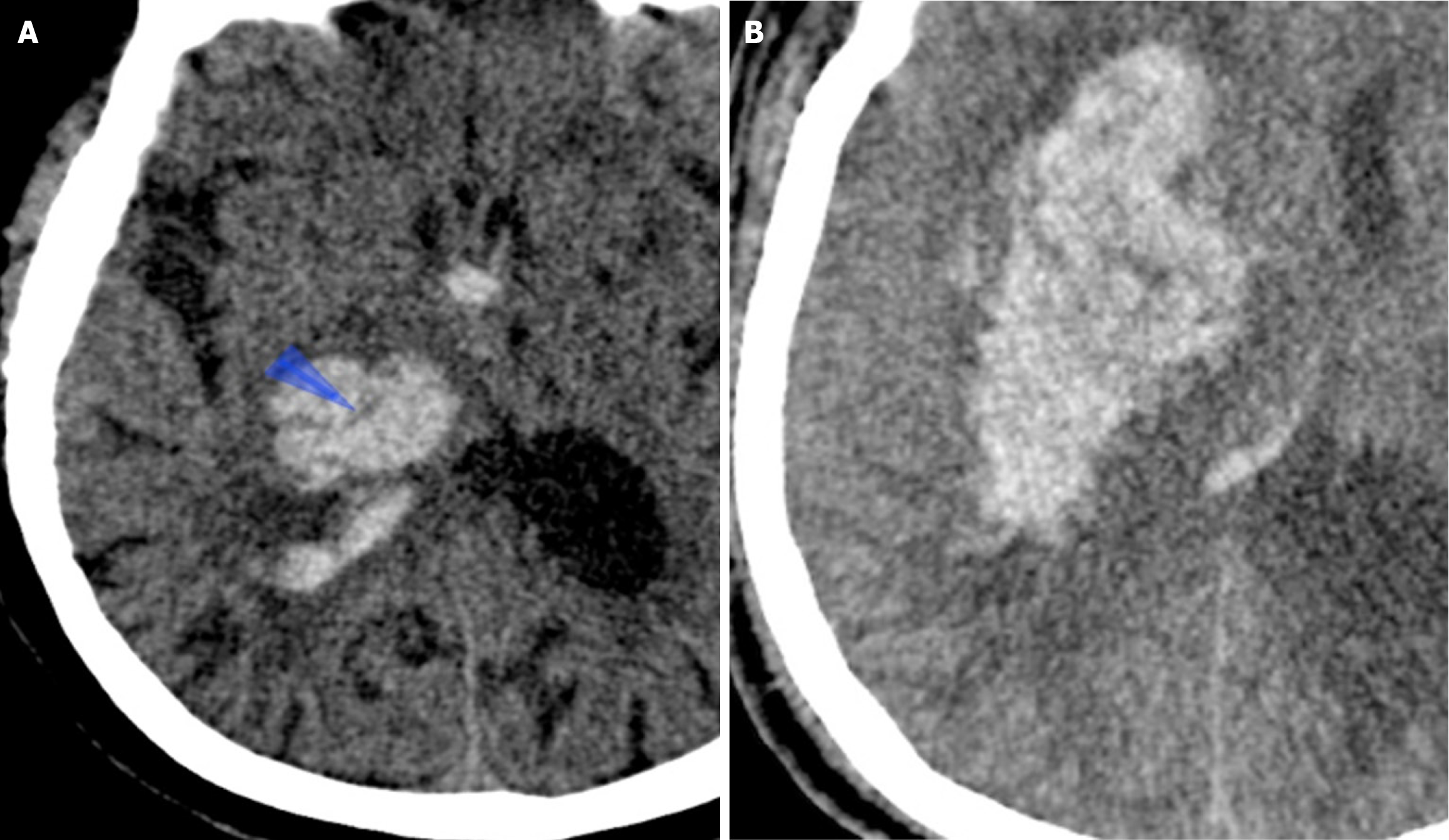
Figure 3 Swirl sign in a 59-year-old man who presented with sudden onset of left-sided weakness.
A: Axial non-contrast computed tomography (NCCT) image performed 1.5 hours after hospital admission showed a right basal ganglia bleed with a swirl sign and intraventricular hemorrhage (blue arrowhead); B: Follow-up NCCT 6-hours later showed a significant hematoma expansion.
- Citation: Parry AH, Fatima SA, Wani M, Jehangir M, Farooq O, Wagay MI, Ashraf O, Hakeem AA. Predicting hematoma growth in spontaneous intracerebral hemorrhage: A simplified non-contrast computed tomography based five-point grading approach. World J Radiol 2025; 17(9): 112983
- URL: https://www.wjgnet.com/1949-8470/full/v17/i9/112983.htm
- DOI: https://dx.doi.org/10.4329/wjr.v17.i9.112983