
















©The Author(s) 2025.
World J Radiol. Sep 28, 2025; 17(9): 112983
Published online Sep 28, 2025. doi: 10.4329/wjr.v17.i9.112983
Published online Sep 28, 2025. doi: 10.4329/wjr.v17.i9.112983
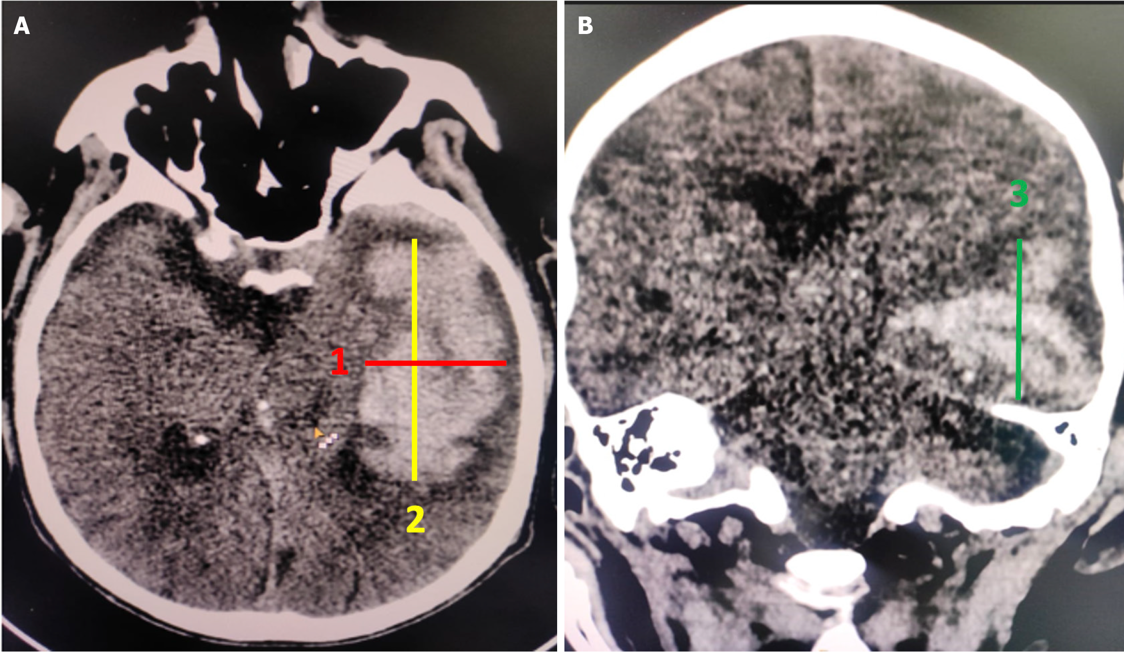
Figure 2 Calculation of volume of intracerebral hematoma on non-contrast computed tomography scan of brain using ABC/2.
A: Maximum width (red line) and maximum length (yellow line) of hematoma in cm; B: Longest cranio-caudal measurement of hematoma on the coronal section. Multiplying the three dimensions and dividing it by 2 yields the final volume in milliliters.
- Citation: Parry AH, Fatima SA, Wani M, Jehangir M, Farooq O, Wagay MI, Ashraf O, Hakeem AA. Predicting hematoma growth in spontaneous intracerebral hemorrhage: A simplified non-contrast computed tomography based five-point grading approach. World J Radiol 2025; 17(9): 112983
- URL: https://www.wjgnet.com/1949-8470/full/v17/i9/112983.htm
- DOI: https://dx.doi.org/10.4329/wjr.v17.i9.112983