
















©The Author(s) 2025.
World J Radiol. Sep 28, 2025; 17(9): 110906
Published online Sep 28, 2025. doi: 10.4329/wjr.v17.i9.110906
Published online Sep 28, 2025. doi: 10.4329/wjr.v17.i9.110906
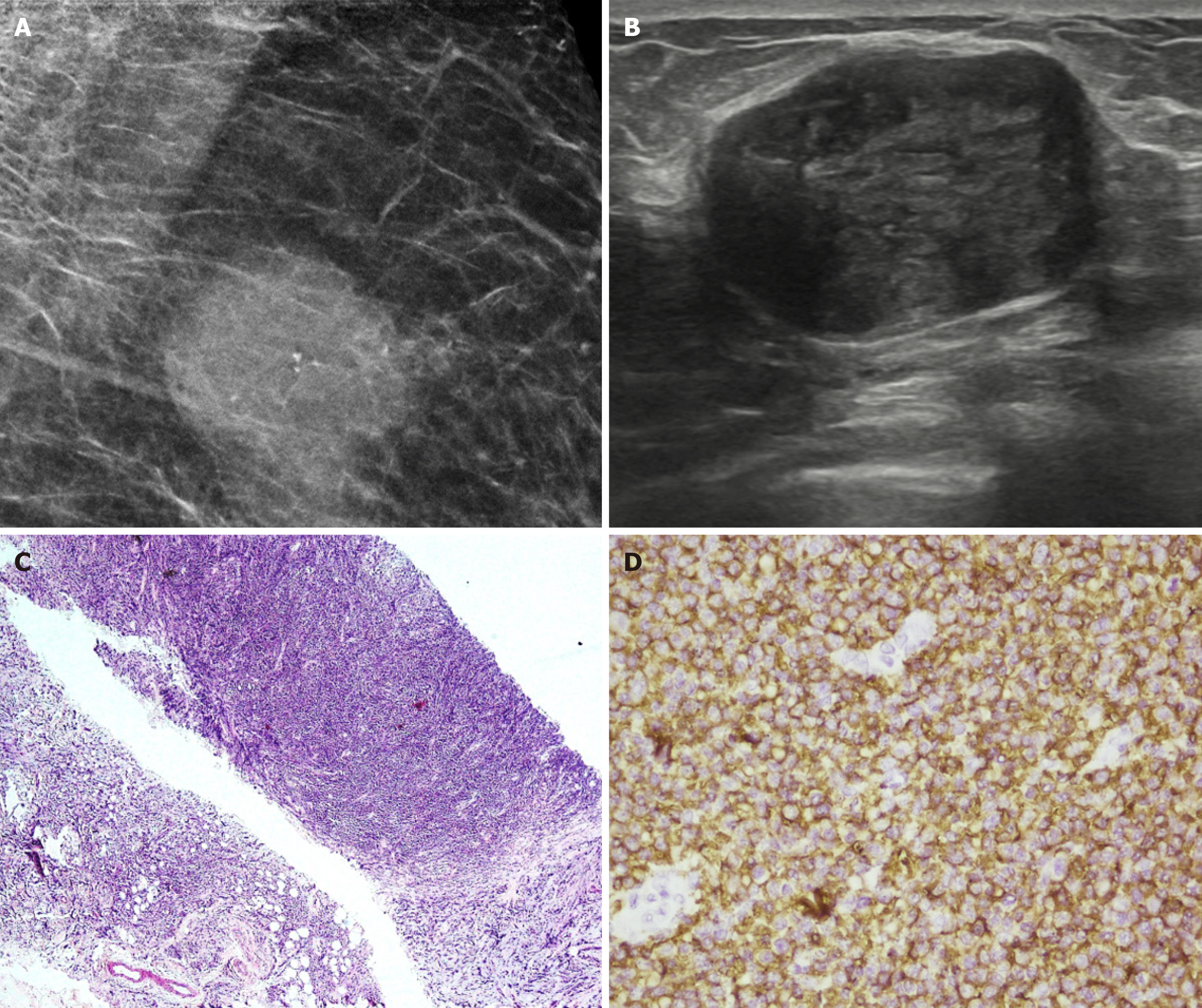
Figure 22 Lymphoma in a 43-year-old male after 3 cycles of chemotherapy.
A: Magnified mediolateral oblique view of the right breast showing a round high-density mass with partially indistinct margins and few punctate calcifications. B: Ultrasound image showing an oval hypoechoic parallel mass with circumscribed margins. C: Histopathology (hematoxylin and eosin, × 40). D: CD20 immunohistochemical staining revealed diffuse large B-cell lymphoma with sheets of atypical lymphoid cells that were positive for CD20.
- Citation: Singla V, Bhatia H, Garg D, Bal A, Sekar A. A compendium of male breast imaging: The road less traveled. World J Radiol 2025; 17(9): 110906
- URL: https://www.wjgnet.com/1949-8470/full/v17/i9/110906.htm
- DOI: https://dx.doi.org/10.4329/wjr.v17.i9.110906