
















©The Author(s) 2025.
World J Radiol. Sep 28, 2025; 17(9): 110906
Published online Sep 28, 2025. doi: 10.4329/wjr.v17.i9.110906
Published online Sep 28, 2025. doi: 10.4329/wjr.v17.i9.110906
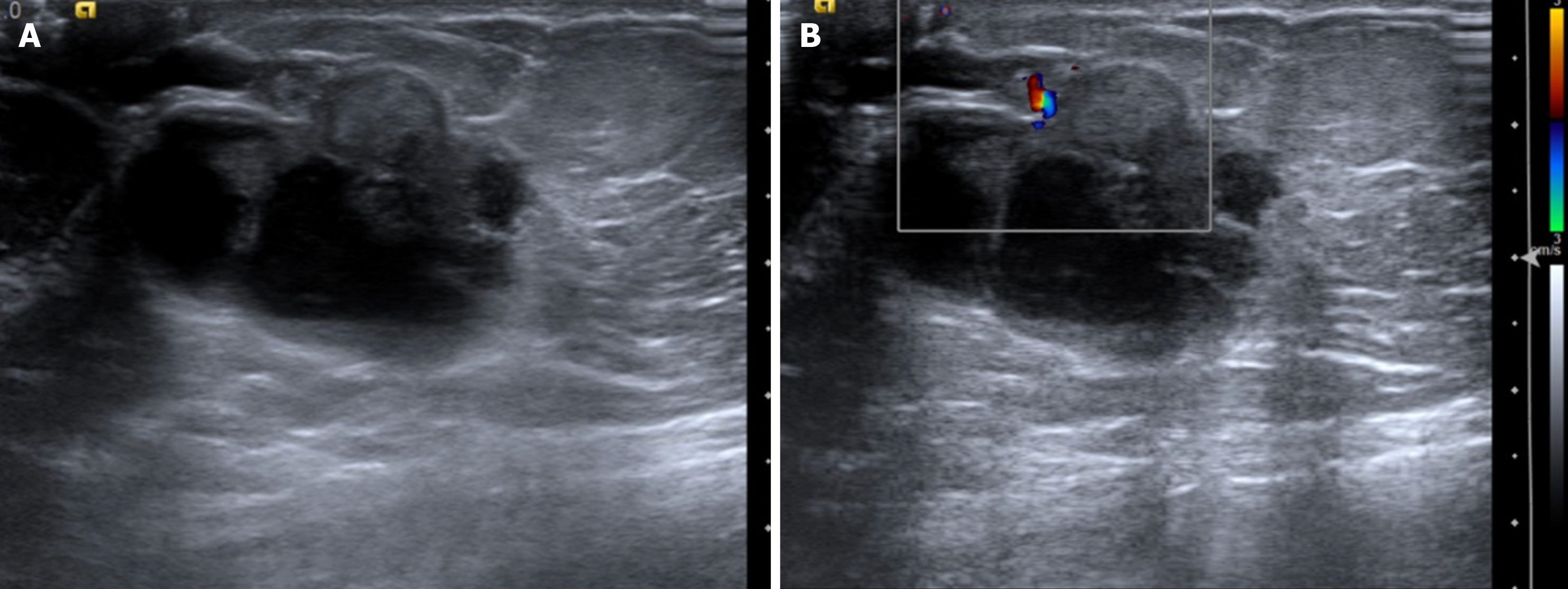
Figure 21 Papillary carcinoma in a 54-year-old male with a hard mass in left breast.
A: Ultrasound; B: Color Doppler images showing an irregular solid-cystic mass with an eccentric solid component, with minimal peripheral vascularity.
- Citation: Singla V, Bhatia H, Garg D, Bal A, Sekar A. A compendium of male breast imaging: The road less traveled. World J Radiol 2025; 17(9): 110906
- URL: https://www.wjgnet.com/1949-8470/full/v17/i9/110906.htm
- DOI: https://dx.doi.org/10.4329/wjr.v17.i9.110906